Copyright
©The Author(s) 2020.
World J Gastroenterol. Nov 21, 2020; 26(43): 6810-6821
Published online Nov 21, 2020. doi: 10.3748/wjg.v26.i43.6810
Published online Nov 21, 2020. doi: 10.3748/wjg.v26.i43.6810
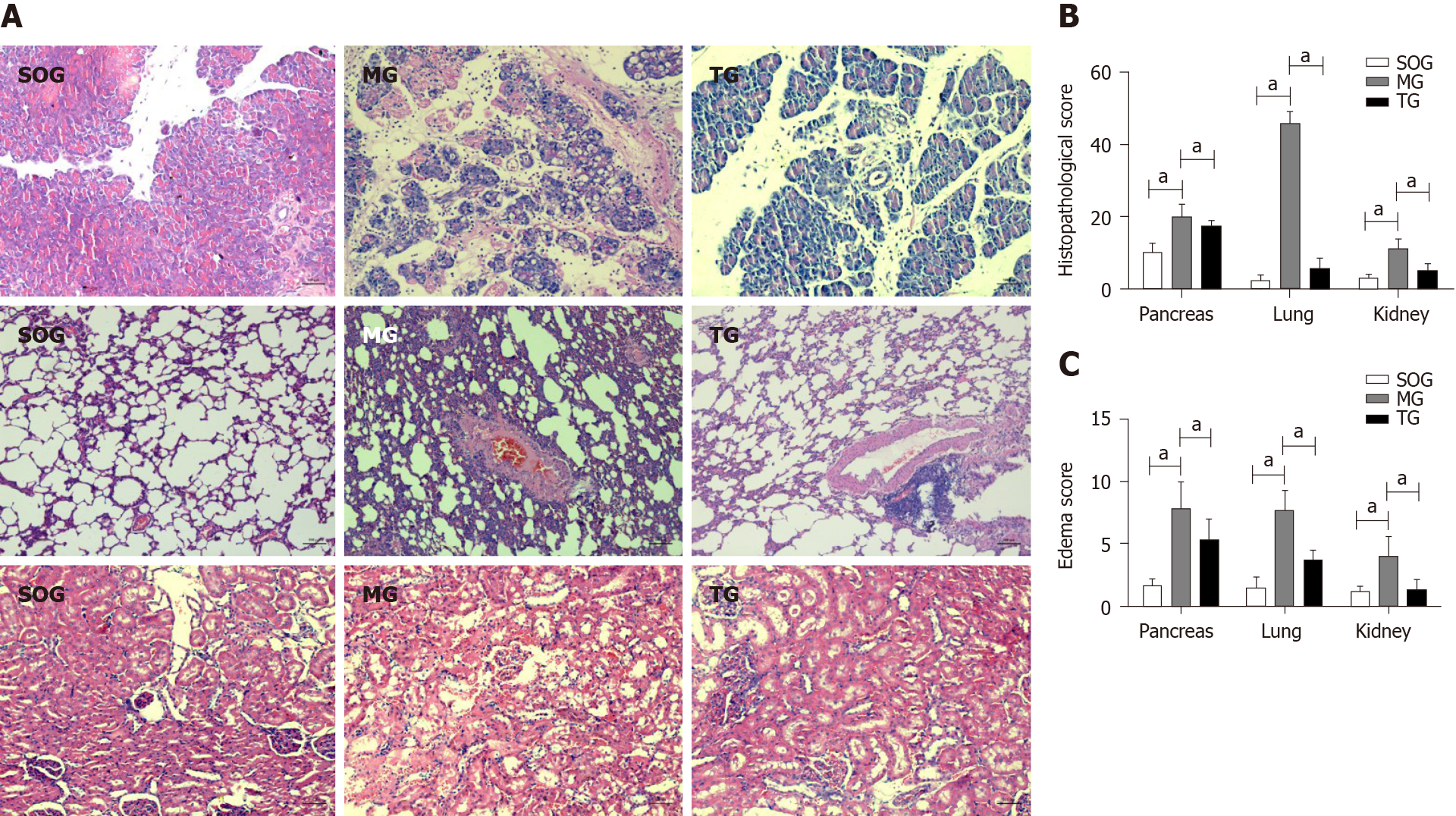
Figure 1 Histological images and pathologic and edema scores of pancreatic, lung, and kidney tissues in the three study groups.
A: Pathological images of the pancreatic, lung, and kidney tissues (hematoxylin-eosin staining, magnification: pancreas and kidney × 200; lung × 100); B: Histological scores of the three types of tissues; C: Edema scores of the three types of tissues. The results are represented as the mean ± SD. aP < 0.05 (n = 12). SOG: Sham operation group; MG: Model group; TG: Treatment group.
Figure 2 Levels of lung injury factors.
A: Levels of superoxide dismutase in three groups; B: Levels of malondialdehyde in three groups. The results are represented as the mean ± SD. aP < 0.05, NS: P > 0.05 (n = 12). SOG: Sham operation group; MG: Model group; TG: Treatment group; SOD: Superoxide dismutase; MDA: Malondialdehyde.
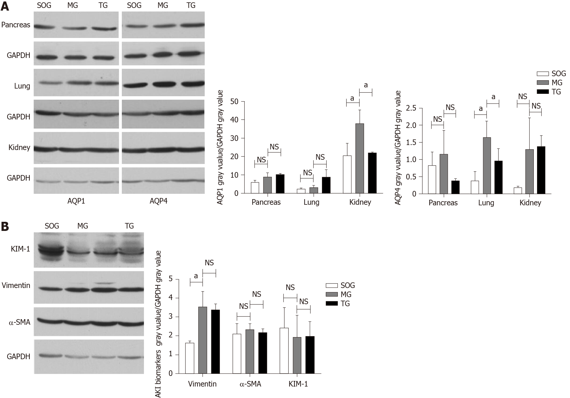
Figure 3 Western blot analysis of expression of aquaporins and acute renal injury biomarkers.
A: Grayscale images and relative expression of aquaporins in three groups; B: Grayscale images and relative expression of acute renal injury biomarkers in three groups. The results are represented as the mean ± SD. aP < 0.05; NS: P > 0.05 (n = 12). SOG: Sham operation group; MG: Model group; TG: Treatment group; MDA: Malondialdehyde; KIM-1: Kidney injury molecule-1; α-SMA: α-smooth muscle actin.
Figure 4 Aquaporins and kidney injury molecule-1 mRNA expression in three groups.
The results are represented as the mean ± SD. aP < 0.05; NS: P > 0.05 (n = 12). MG: Model group; TG: Treatment group; KIM-1: Kidney injury molecule-1; AQP1: Aquaporin 1.
- Citation: Hu J, Zhang YM, Miao YF, Zhu L, Yi XL, Chen H, Yang XJ, Wan MH, Tang WF. Effects of Yue-Bi-Tang on water metabolism in severe acute pancreatitis rats with acute lung-kidney injury. World J Gastroenterol 2020; 26(43): 6810-6821
- URL: https://www.wjgnet.com/1007-9327/full/v26/i43/6810.htm
- DOI: https://dx.doi.org/10.3748/wjg.v26.i43.6810


