Copyright
©The Author(s) 2020.
World J Gastroenterol. Nov 14, 2020; 26(42): 6614-6625
Published online Nov 14, 2020. doi: 10.3748/wjg.v26.i42.6614
Published online Nov 14, 2020. doi: 10.3748/wjg.v26.i42.6614
Figure 1 Magnetic compression anastomosis devices.
A: Traditional nummular magnetic compression anastomosis (MCA) devices of different sizes used in experiment 1; B: Fedora-type MCA devices with different design used in experiment 2; C: Schematic diagram of the fedora-type MCA device.
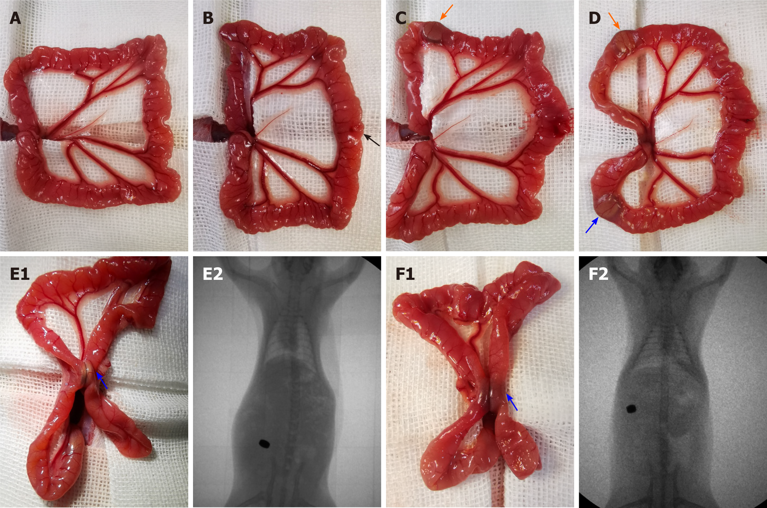
Figure 2 Surgical procedure and X-ray fluoroscopy.
A: The small intestine was removed; B: A 6 mm incision was made 12 cm distal to the cecum (black arrow); C: The daughter part (orange arrow) was inserted; D: The parent part (blue arrow) was inserted; E1: Two magnets of the traditional nummular magnetic compression anastomosis (MCA) device were coupled (blue arrow) to compress the ileac wall; E2: Accurate coupling of the daughter and parent magnets in experiment 1 was confirmed using X-ray; F1: Two parts of the fedora-type MCA device were coupled (blue arrow); F2: Accurate coupling of the daughter and parent parts in experiment 2 was confirmed using X-ray.
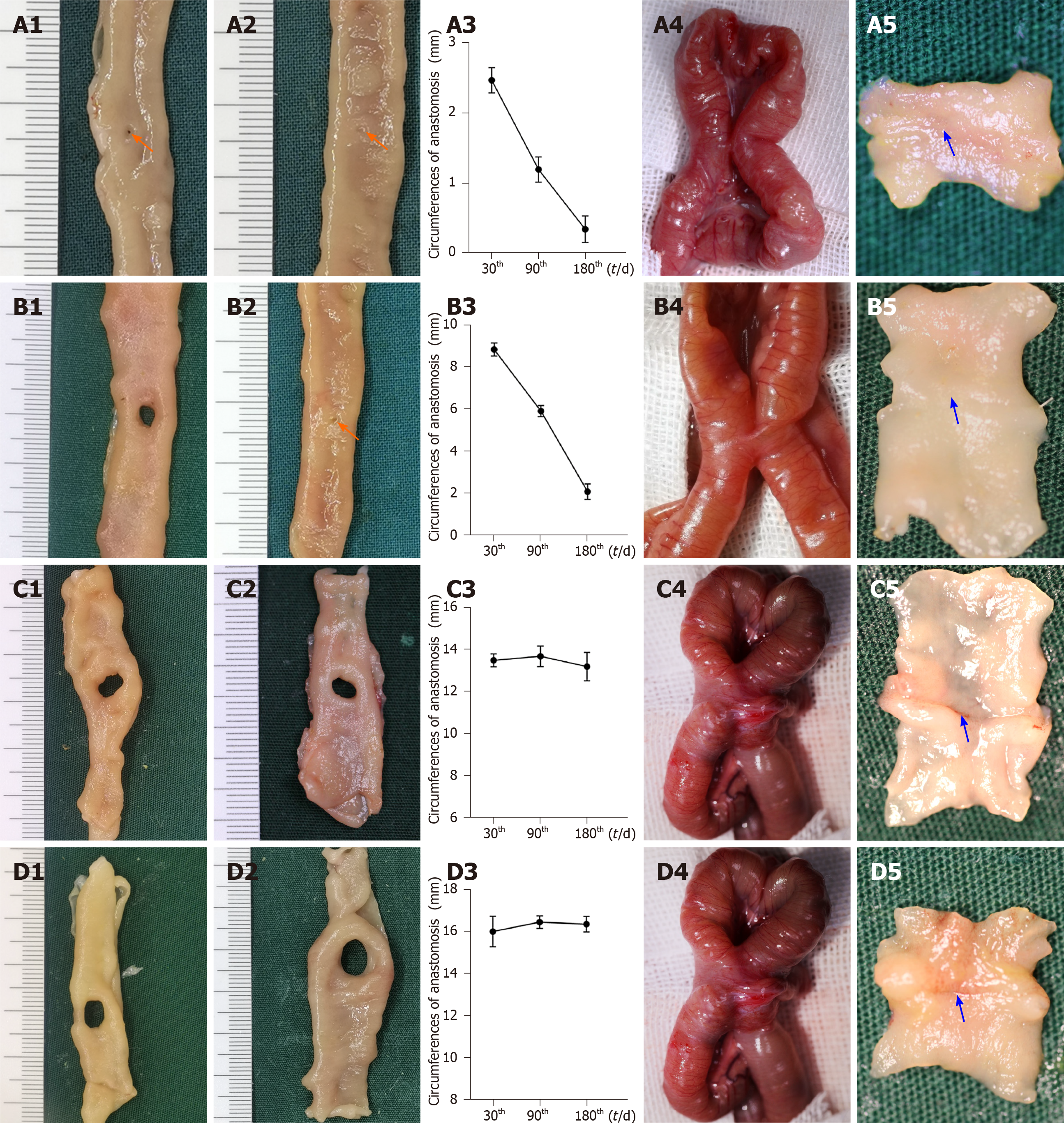
Figure 3 Gross appearance of anastomoses using traditional nummular magnetic compression anastomosis devices.
A: Group 1.1 (Φ3 mm): The size of anastomosis 30 d after magnetic compression anastomosis (MCA) (A1), the size of anastomosis 180 d after MCA (A2), the change in anastomosis circumferences after MCA (A3), serosa side of anastomosis (A4), and mucosa side of anastomosis (A5); B: Group 1.2 (Φ4 mm): The size of anastomosis 30 d after MCA (B1), the size of anastomosis 180 d after MCA (B2), the change in anastomosis circumferences after MCA (B3), serosa side of anastomosis (B4), and mucosa side of anastomosis (B5); C: Group 1.3 (Φ5 mm): The size of anastomosis 30 d after MCA (C1), the size of anastomosis 180 d after MCA (C2), the change in anastomosis circumferences after MCA (C3), serosa side of anastomosis (C4), and mucosa side of anastomosis (C5); D: Group 1.4 (Φ6 mm): The size of anastomosis 30 d after MCA (D1), the size of anastomosis 180 d after MCA (D2), the change in anastomosis circumferences after MCA (D3), serosa side of anastomosis (D4), and mucosa side of anastomosis (D5). Orange arrows: Anastomosis; blue arrows: Anastomotic line.
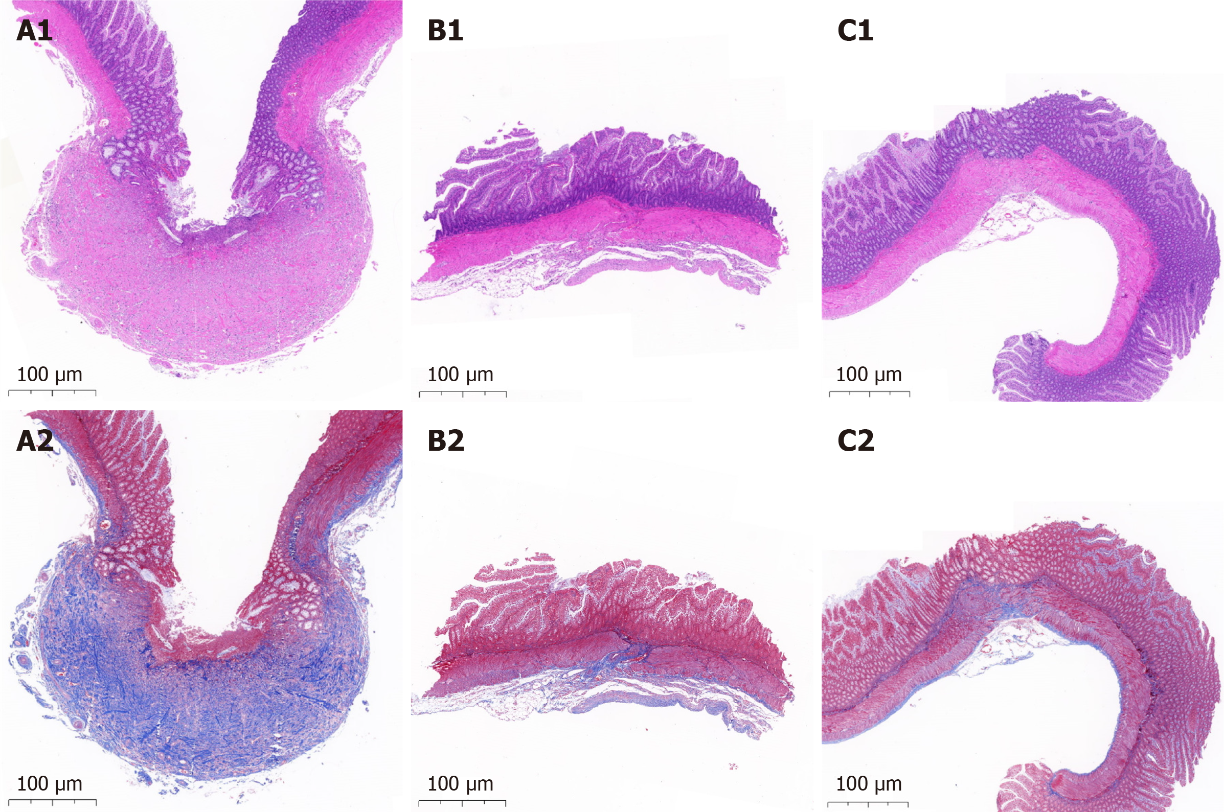
Figure 4 Microscopic appearance of anastomosis.
A: Larger size groups of traditional nummular magnetic compression anastomosis (MCA) devices (Group 1.3 and 1.4): Hematoxylin and eosin staining (A1) and Masson’s trichrome staining (A2); B: Smaller size groups of traditional nummular MCA devices (Group 1.1 and 1.2): Hematoxylin and eosin staining (B1) and Masson’s trichrome staining (B2); C: Fedora-type MCA device: Hematoxylin and eosin staining (C1) and Masson’s trichrome staining (C2).
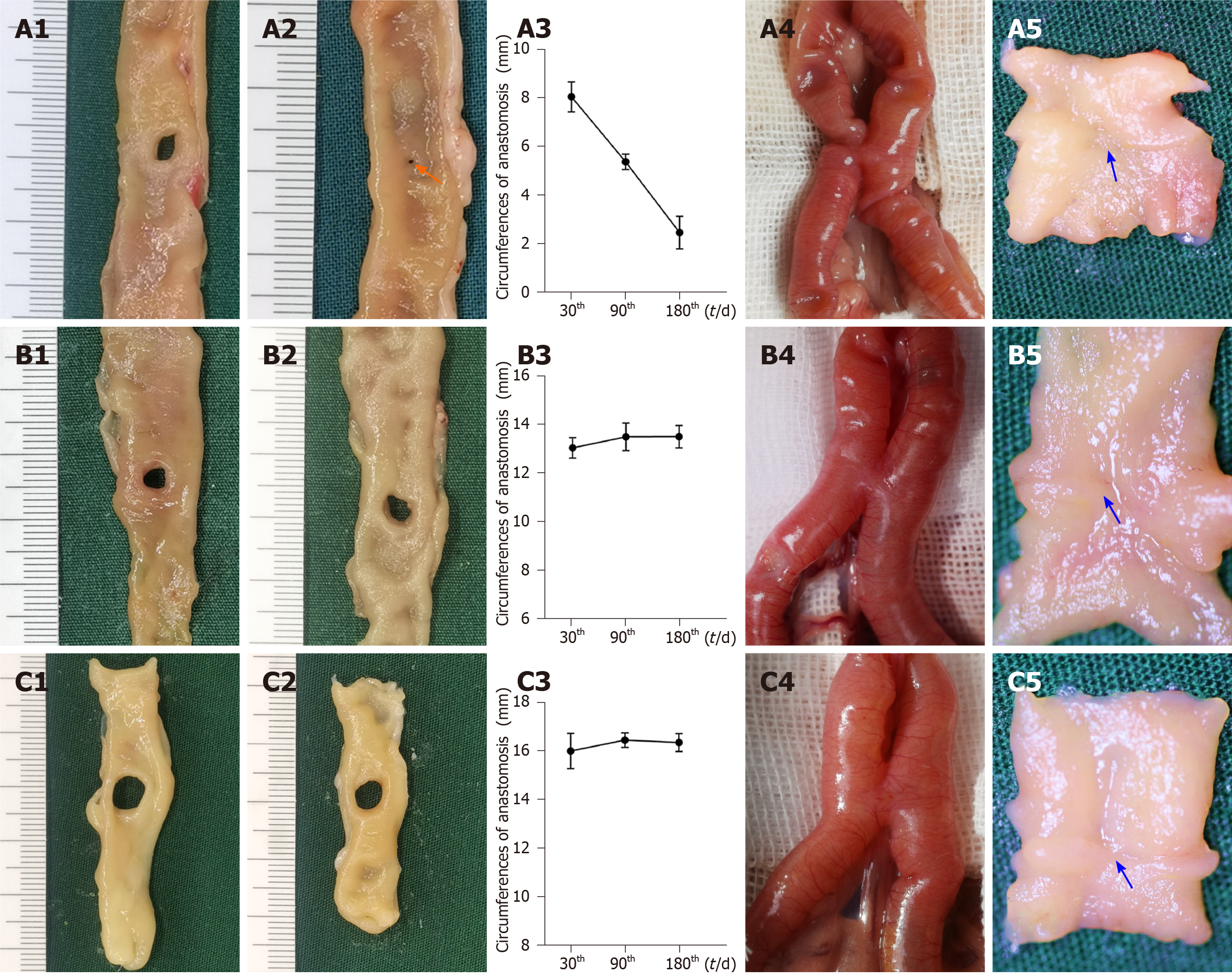
Figure 5 Gross appearance of anastomoses using the fedora-type magnetic compression anastomosis devices.
A: Group 2.1 (with a Φ4 mm sheet metal): The size of anastomosis 30 d after magnetic compression anastomosis (MCA) (A1), the size of anastomosis 180 d after MCA (A2), the change in anastomosis circumferences after MCA (A3), serosa side of anastomosis (A4), and mucosa side of anastomosis (A5); B: Group 2.2 (with a Φ5 mm sheet metal): The size of anastomosis 30 d after MCA (B1), the size of anastomosis 180 d after MCA (B2), the change in anastomosis circumferences after MCA (B3), serosa side of anastomosis (B4), and mucosa side of anastomosis (B5); C: Group 2.3 (with a Φ6 mm sheet metal): The size of anastomosis 30 d after MCA (C1), the size of anastomosis 180 d after MCA (C2), the change in anastomosis circumferences after MCA (C3), serosa side of anastomosis (C4), and mucosa side of anastomosis (C5). Orange arrows: Anastomosis; blue arrows: Anastomotic line.
- Citation: Chen H, Ma T, Wang Y, Zhu HY, Feng Z, Wu RQ, Lv Y, Dong DH. Fedora-type magnetic compression anastomosis device for intestinal anastomosis. World J Gastroenterol 2020; 26(42): 6614-6625
- URL: https://www.wjgnet.com/1007-9327/full/v26/i42/6614.htm
- DOI: https://dx.doi.org/10.3748/wjg.v26.i42.6614


