Copyright
©The Author(s) 2020.
World J Gastroenterol. Sep 7, 2020; 26(33): 5050-5059
Published online Sep 7, 2020. doi: 10.3748/wjg.v26.i33.5050
Published online Sep 7, 2020. doi: 10.3748/wjg.v26.i33.5050
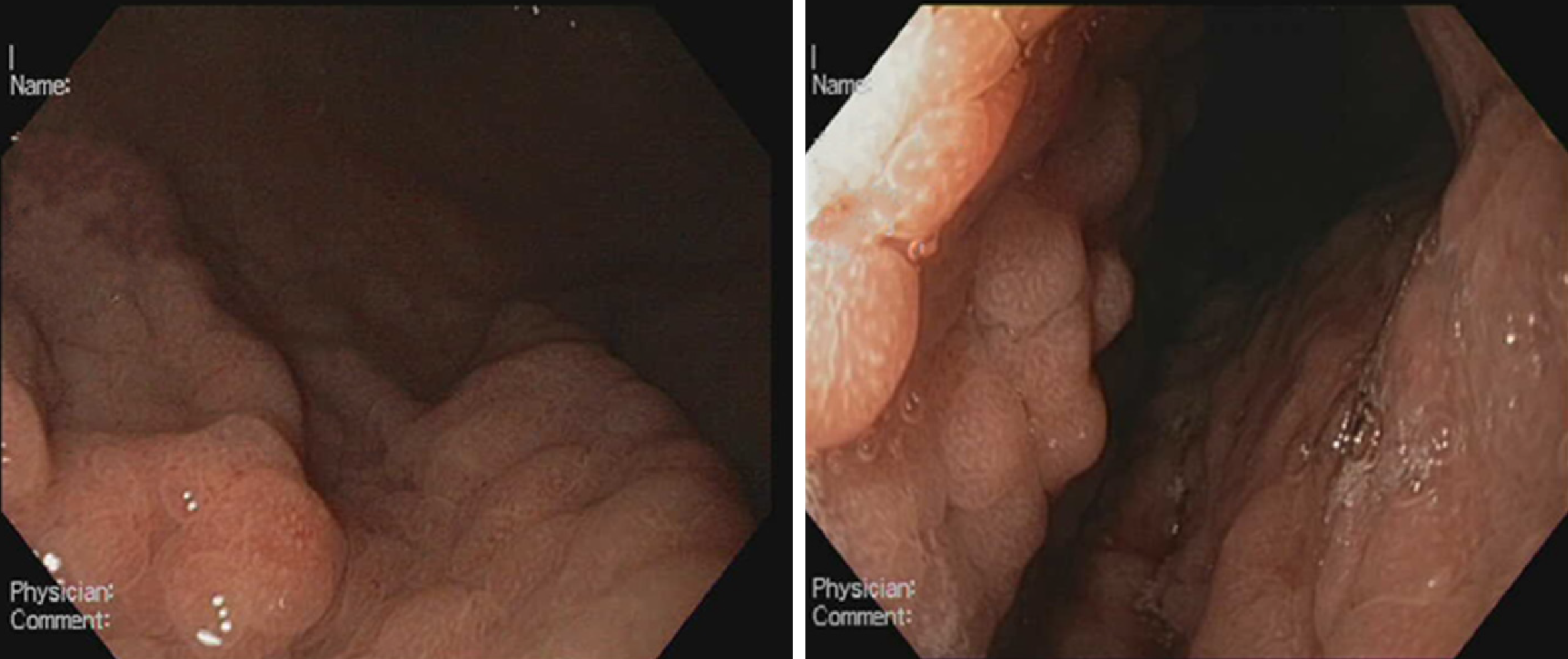
Figure 1 Endoscopic appearance of Helicobacter pylori-negative Russell body gastritis.
Figure 2 Russell body gastritis of the fundus mucosa in the standard histological stain Hematoxylin-eosin, × 400.
A: The initial biopsy specimen shows abundant plasma cell inflammatory infiltrate, rich in Russell body and mature plasma cell; B: The follow-up biopsy revealed no change in plasma cell inflammatory infiltrate, but with decreased distribution Russell body and mature plasma cells; C: Third biopsy, twelve months after the initial diagnosis, demonstrated that chronic inflammatory infiltrate in the fundus mucosa is less pronounced, rich in plasma cells, with almost absent Russell body and mature plasma cells.
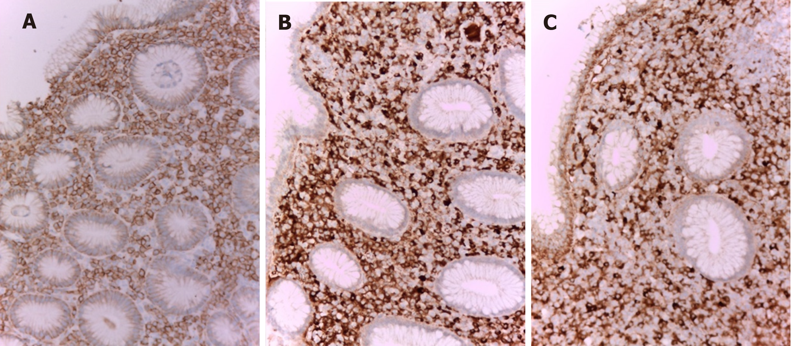
Figure 3 Immunohistochemical stains in Russell body gastritis (initial biopsy) × 200.
A: The inflammatory infiltrate in the gastric fundus mucosa is CD79a positive, which is in support of its homogeneous plasmocytic nature; B: Kappa; C: Lambda. The plasma cells are polyclonal both kappa (B) and lambda (C) light chains are positive.
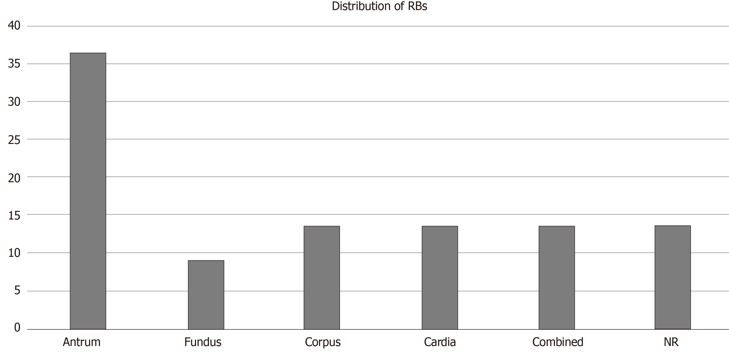
Figure 4 Distribution of Russell bodies in the stomach: in all cases from the literature.
RB: Russell body.
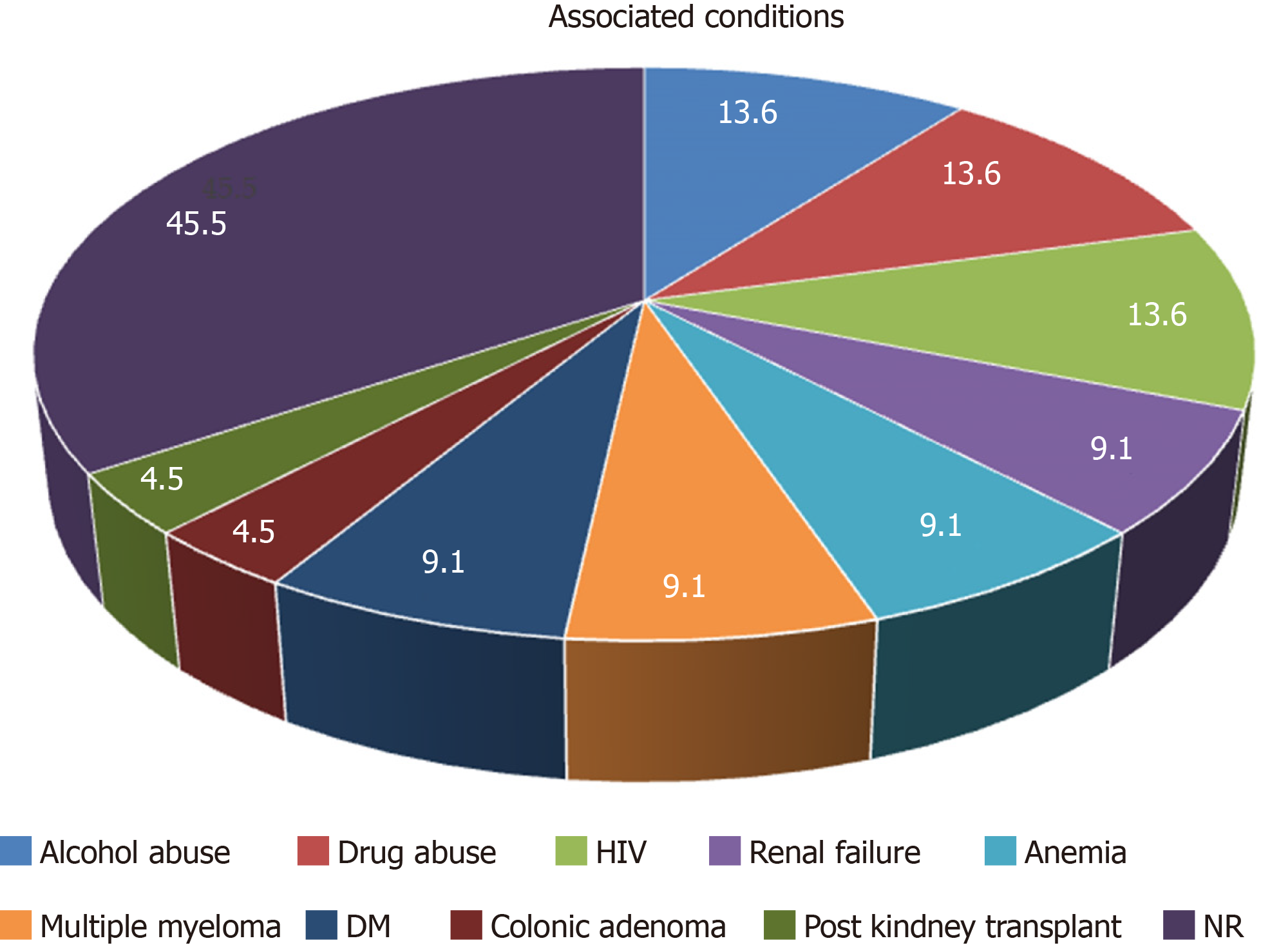
Figure 5 Associated conditions in patients with Helicobacter pylori-negative Russell body gastritis according to the available literature.
- Citation: Peruhova M, Peshevska-Sekulovska M, Georgieva V, Panayotova G, Dikov D. Surveilling Russell body Helicobacter pylori-negative gastritis: A case report and review of literature. World J Gastroenterol 2020; 26(33): 5050-5059
- URL: https://www.wjgnet.com/1007-9327/full/v26/i33/5050.htm
- DOI: https://dx.doi.org/10.3748/wjg.v26.i33.5050


