Copyright
©The Author(s) 2020.
World J Gastroenterol. Aug 7, 2020; 26(29): 4372-4377
Published online Aug 7, 2020. doi: 10.3748/wjg.v26.i29.4372
Published online Aug 7, 2020. doi: 10.3748/wjg.v26.i29.4372
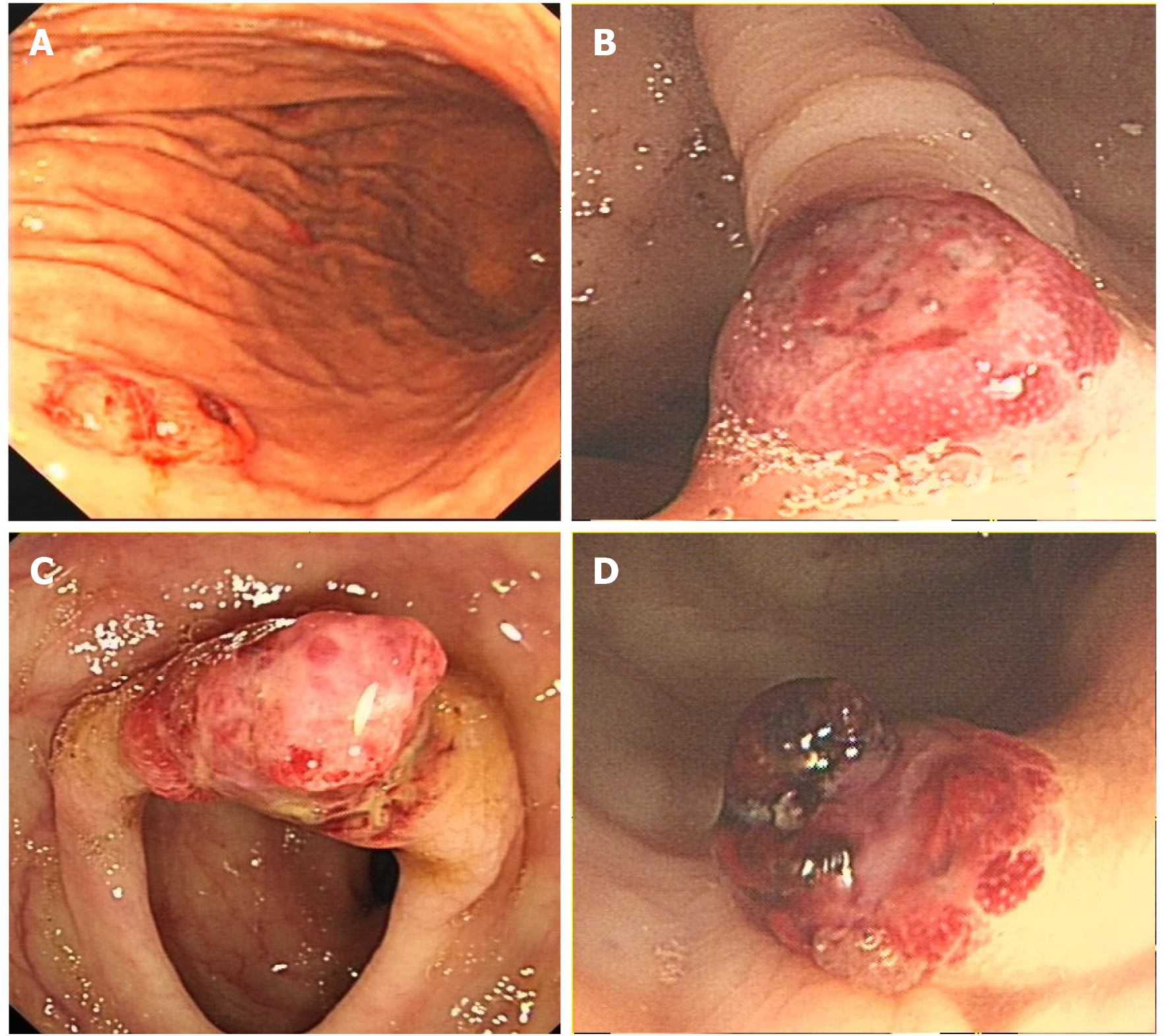
Figure 1 Endoscopy.
A: Gastroscopy revealed a 0.5 cm × 0.6 cm centrally ulcerated polypoid lesion in the gastric body; B: A 0.8 cm × 0.8 cm polypoid lesion in the ascending colon; C: A 1.2 cm × 1.0 cm hyperaemic mass in the transverse colon; and D: A 0.8 cm × 0.8 cm polypoid nodule with hemorrhagic tendency and blood clots in the sigmoid colon.
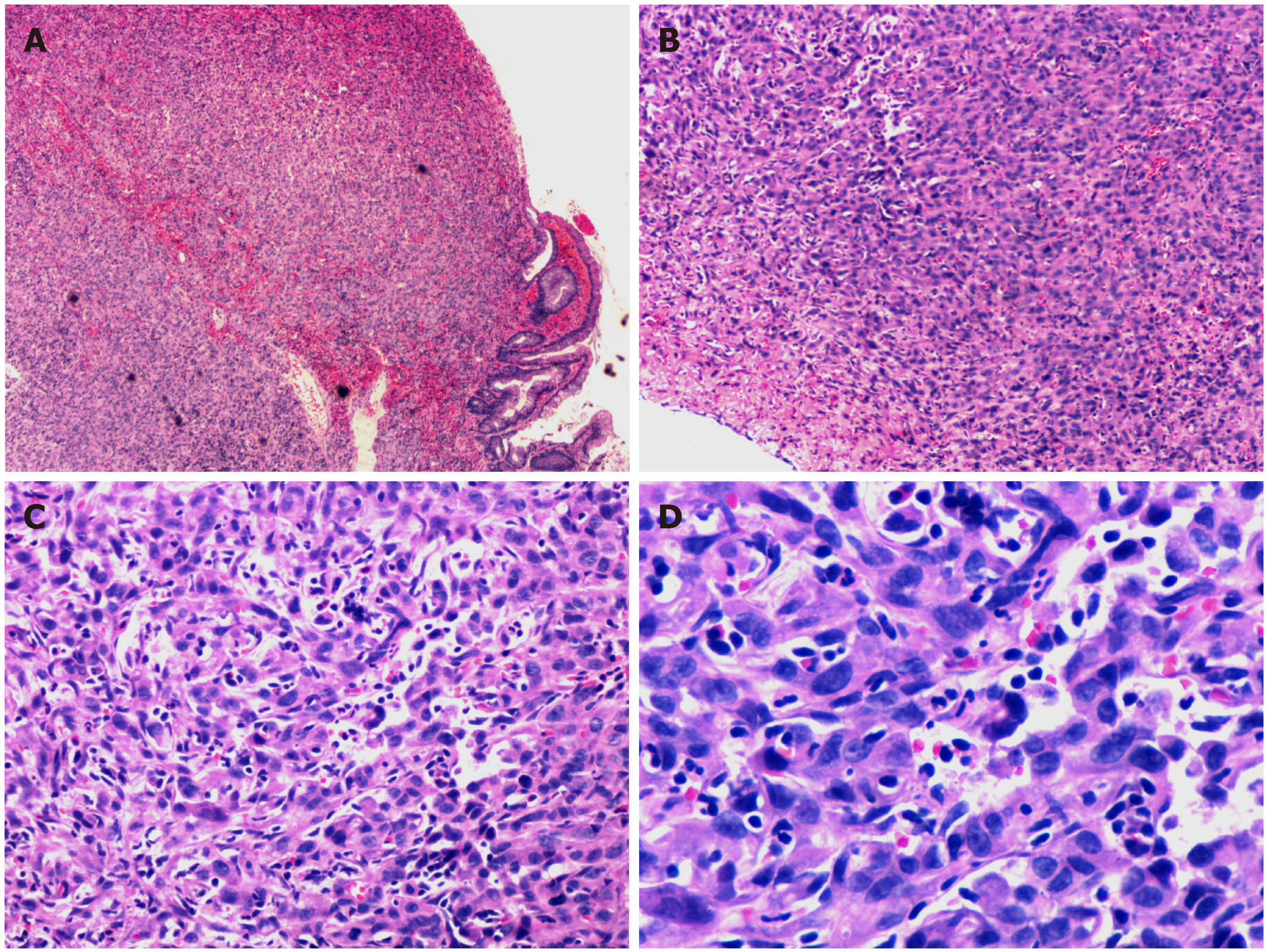
Figure 2 Histopathological findings.
Poorly differentiated neoplasms consisting of large cells with pleomorphic nuclei and abundant eosinophilic cytoplasm were observed. A: Haematoxylin and eosin (HE) staining section × 40; B: HE staining section (× 100); C: HE staining section (× 200); and D: HE staining section (× 400).
Figure 3 Immunohistochemical staining.
A: Immunostaining for CD31; B: Immunostaining for CD34; and C: Immunostaining for pan-cytokeratin.
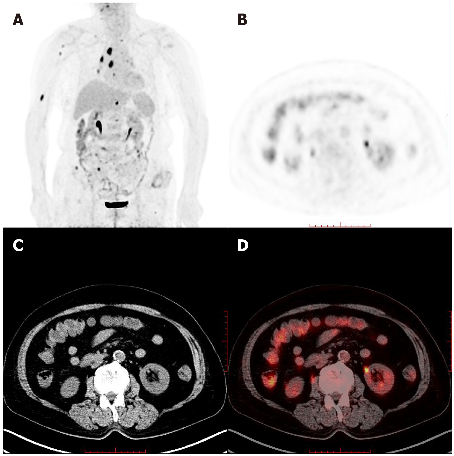
Figure 4 Whole-body maximum intensity projection 18F- fluorodeoxyglucose and positron emission tomography image.
A: Remarkably increased fluorodeoxyglucose metabolism in the colon and mediastinal lymph nodes, as well as the right humerus; B: Positron emission tomography; C: Computed tomography; and D: Positron emission tomography/computed tomography in axial projection showed multiple tubular hypermetabolic lesions in the colon.
- Citation: Chen YW, Dong J, Chen WY, Dai YN. Multifocal gastrointestinal epithelioid angiosarcomas diagnosed by endoscopic mucosal resection: A case report. World J Gastroenterol 2020; 26(29): 4372-4377
- URL: https://www.wjgnet.com/1007-9327/full/v26/i29/4372.htm
- DOI: https://dx.doi.org/10.3748/wjg.v26.i29.4372


