Copyright
©The Author(s) 2020.
World J Gastroenterol. Jan 7, 2020; 26(1): 55-69
Published online Jan 7, 2020. doi: 10.3748/wjg.v26.i1.55
Published online Jan 7, 2020. doi: 10.3748/wjg.v26.i1.55
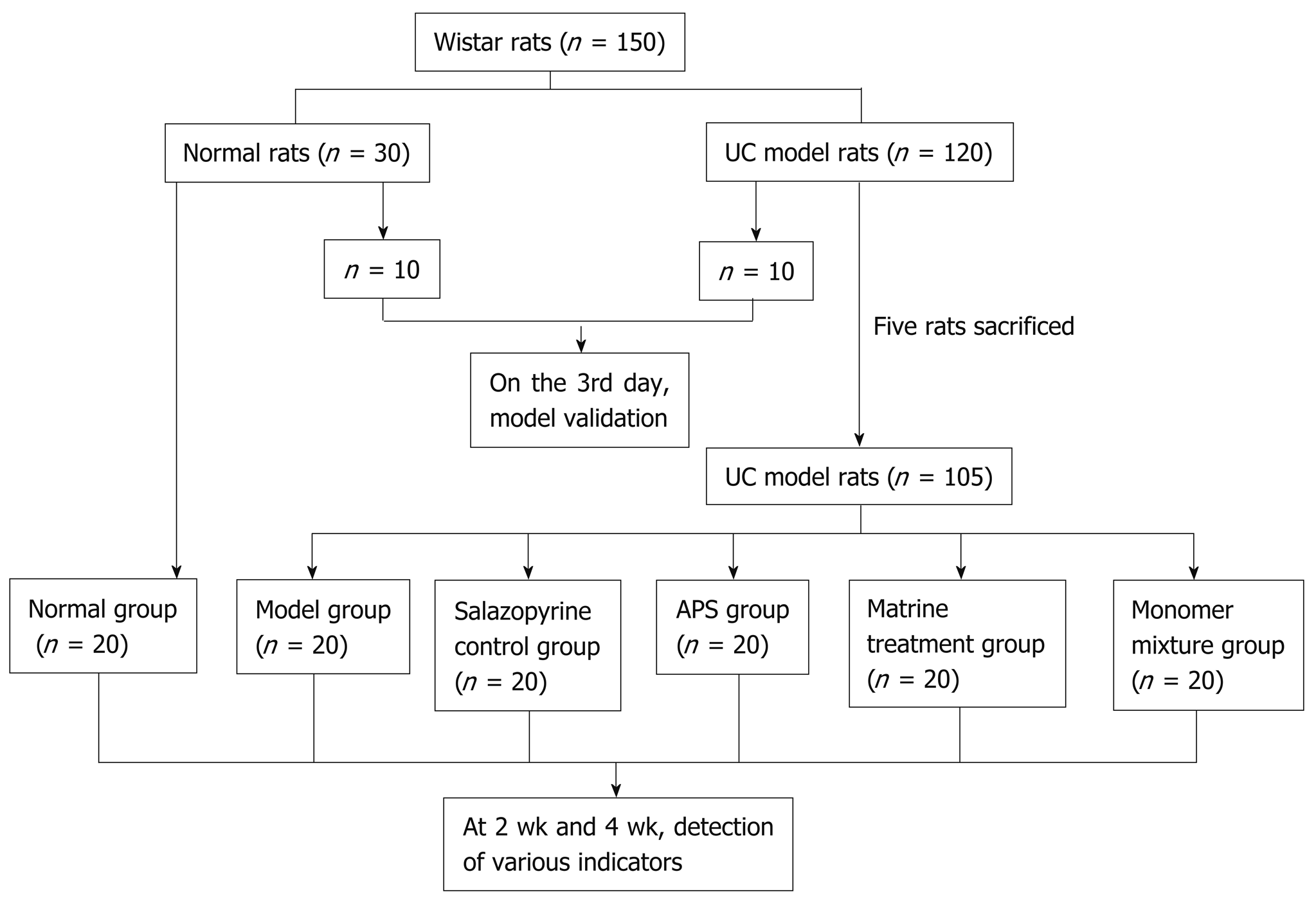
Figure 1 Flow chart of animal grouping.
APS: Astragalus polysaccharides.
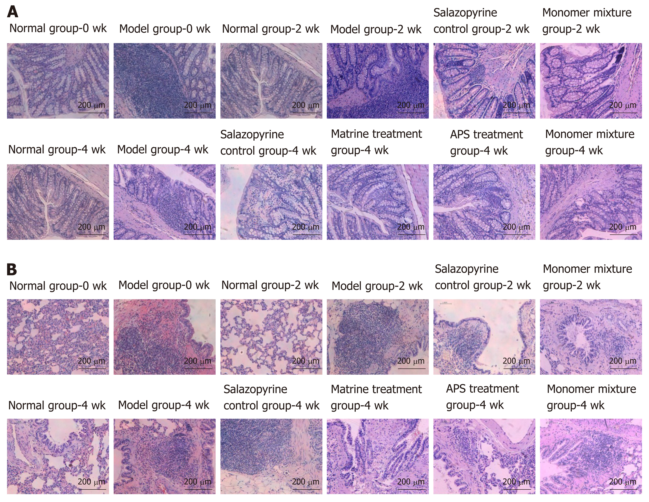
Figure 2 Astragalus polysaccharides combined with matrine improve histopathological changes in rats with ulcerative colitis.
A and B: Histopathological changes of colon (A) and lung tissues (B) in various groups analyzed by hematoxylin-eosin staining. APS: Astragalus polysaccharides.
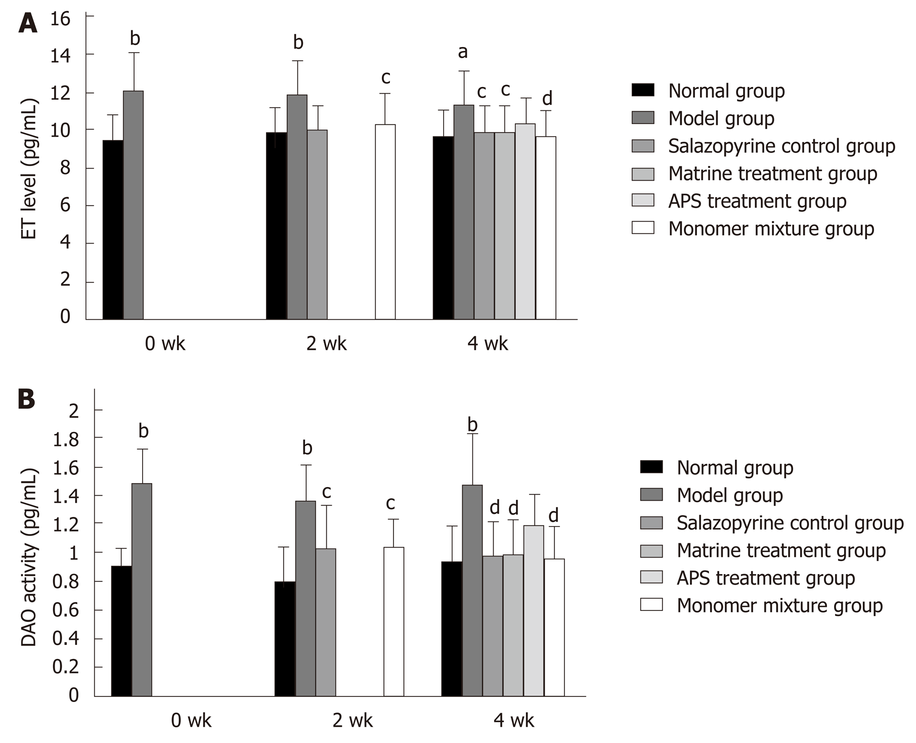
Figure 3 Astragalus polysaccharides combined with matrine inhibit diamine oxidase activity in rats with ulcerative colitis.
A: Serum endotoxin levels in various groups detected by enzyme-linked immunosorbent assay; B: Serum diamine oxidase activity in various groups detected by spectrophotometry. aP < 0.05 and bP < 0.01 vs normal group; cP < 0.05 and dP < 0.01 vs model group. APS: Astragalus polysaccharides; ET: Endotoxin; DAO: Diamine oxidase.
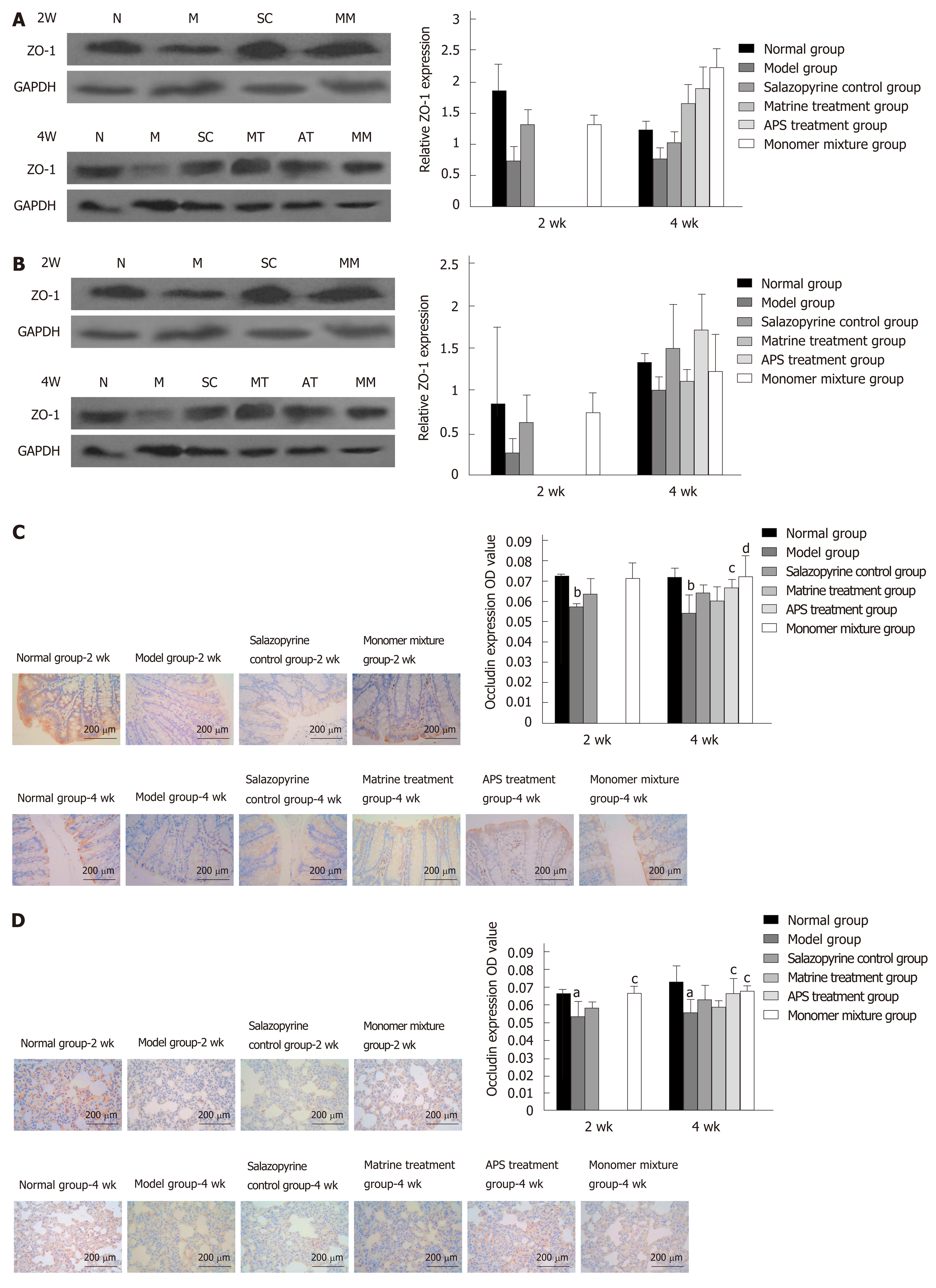
Figure 4 Astragalus polysaccharides combined with matrine inhibit the expression of zonula occludens-1 and Occludin in lung and colon tissues of rats with ulcerative colitis.
A and B: Zonula occludens-1 (ZO-1) expression in colon (A) and lung tissues (B) in various groups detected by Western blot analysis; C and D: Occludin expression in colon (C) and lung tissues (D) in various groups detected by immunohistochemistry analysis. aP < 0.05 and bP < 0.01 vs normal group; cP < 0.05 and dP < 0.01 vs model group. APS: Astragalus polysaccharides; ZO-1: Zonula occludens-1; N: Normal group; M: Model group; SC: Salazopyrine control group; MT: Matrine treatment group; AT: Astragalus polysaccharides treatment group; MM: Monomer mixture group.
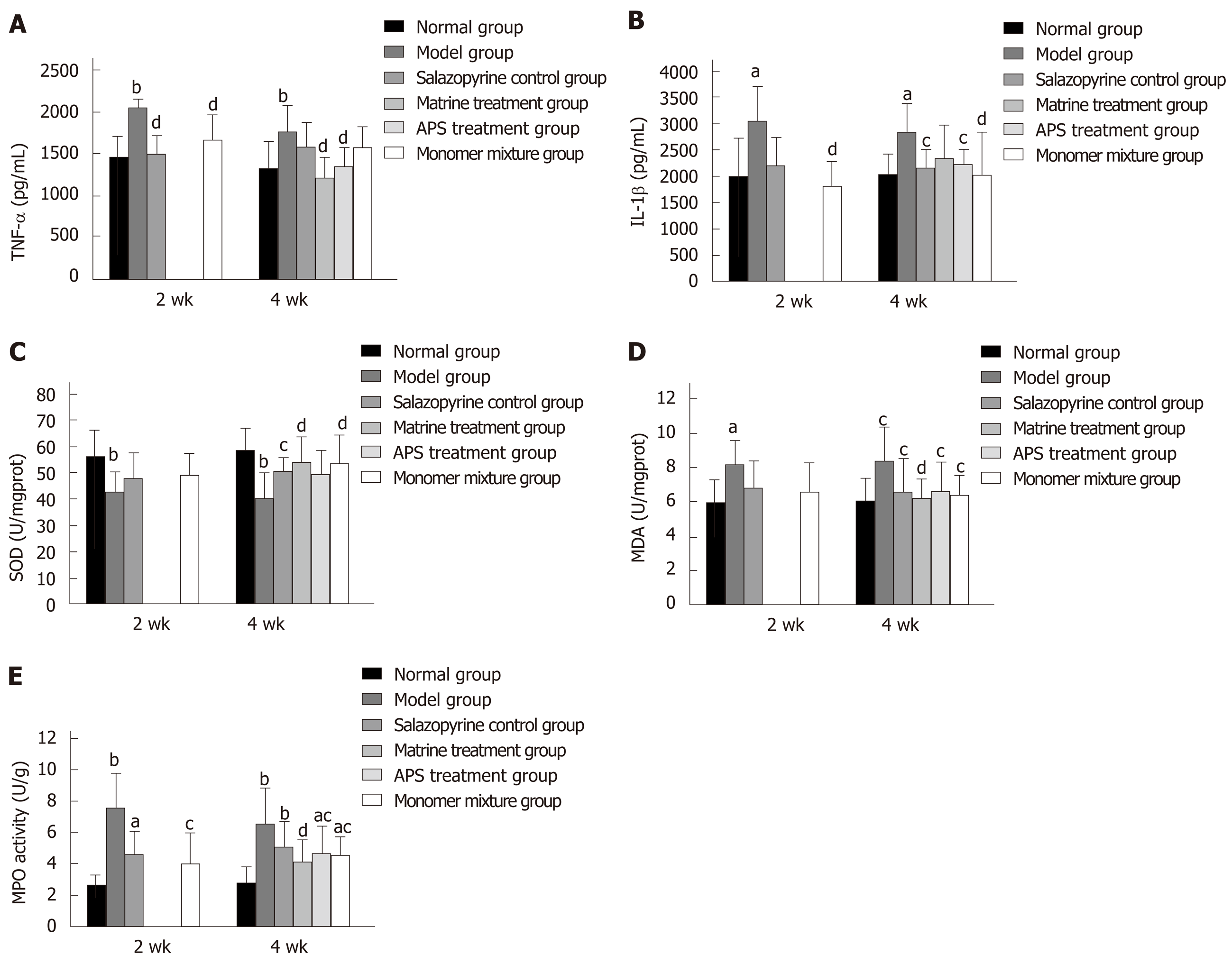
Figure 5 Astragalus polysaccharides combined with matrine relieve inflammatory response and oxidative stress injury in lung tissues of rats with ulcerative colitis.
A and B: Tumor necrosis factor-α (A) and interleukin-1β levels (B) in lung tissues in various groups detected by enzyme-linked immunosorbent assay (ELISA); C-E: Activities of superoxide dismutase (C), malondialdehyde (D), and myeloperoxidase (E) in lung tissues in various groups determined with commercial detection kits. aP < 0.05 and bP < 0.01 vs normal group; cP < 0.05 and dP < 0.01 vs model group. APS: Astragalus polysaccharides; TNF-α: Tumor necrosis factor-α; IL-1β: Interleukins-1β; MPO: Myeloperoxidase.
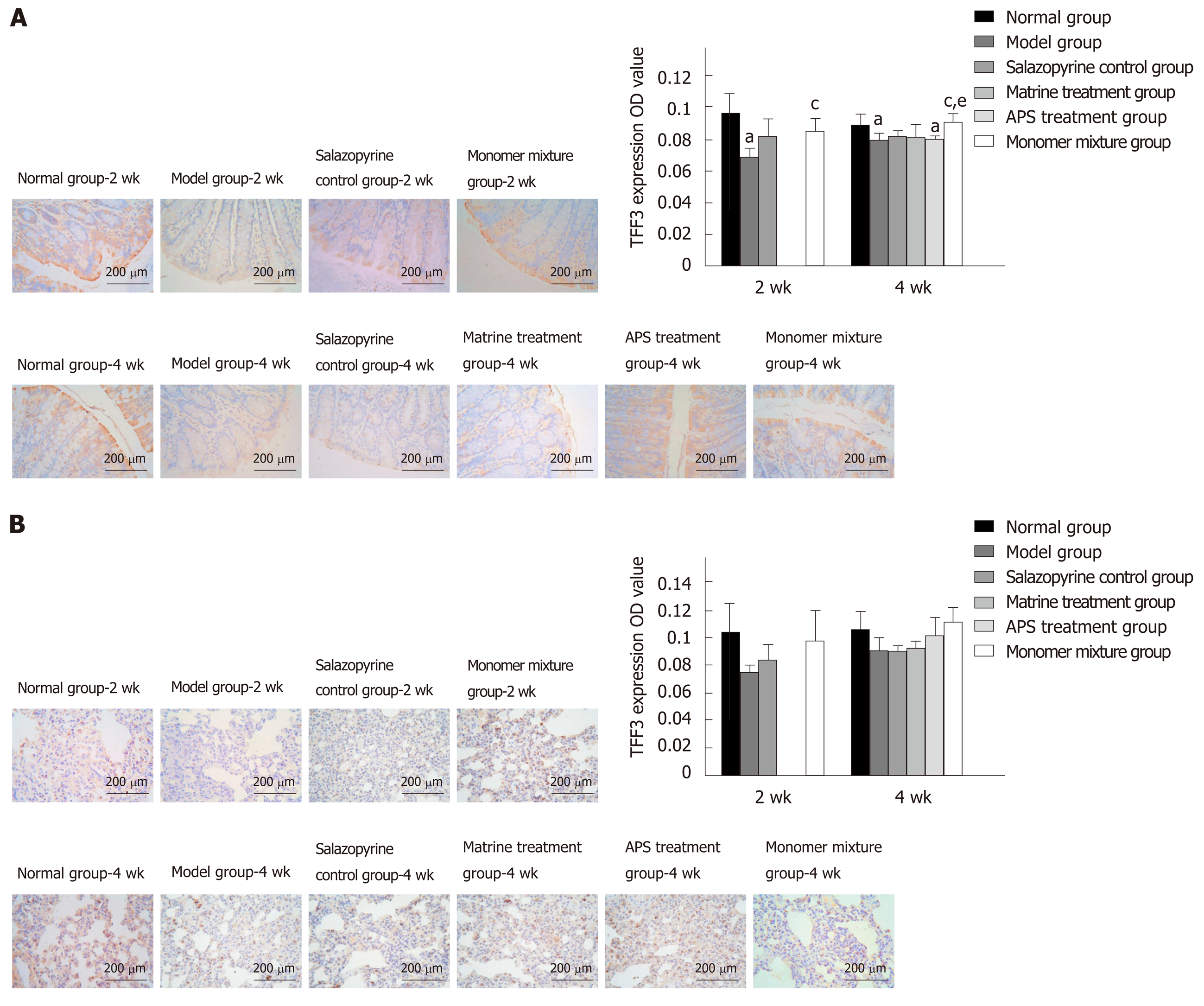
Figure 6 Astragalus polysaccharides combined with matrine increase trefoil factor 3 expression in lung and colon tissues of rats with ulcerative colitis.
A and B: Trefoil factor 3 (TFF3) expression in colon (A) and lung tissues (B) in various groups detected by immunohistochemistry analysis. aP < 0.05 vs normal group; cP < 0.05 vs model group; eP < 0.05 vs salazopyrine control group. APS: Astragalus polysaccharides; TFF3: Trefoil factor 3.
- Citation: Yan X, Lu QG, Zeng L, Li XH, Liu Y, Du XF, Bai GM. Synergistic protection of astragalus polysaccharides and matrine against ulcerative colitis and associated lung injury in rats. World J Gastroenterol 2020; 26(1): 55-69
- URL: https://www.wjgnet.com/1007-9327/full/v26/i1/55.htm
- DOI: https://dx.doi.org/10.3748/wjg.v26.i1.55


