Copyright
©The Author(s) 2019.
World J Gastroenterol. Feb 21, 2019; 25(7): 848-858
Published online Feb 21, 2019. doi: 10.3748/wjg.v25.i7.848
Published online Feb 21, 2019. doi: 10.3748/wjg.v25.i7.848
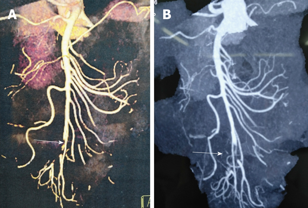
Figure 1 Computed tomography angiography images.
A and B: Acute embolic occlusion of the superior mesenteric artery indicated by the white arrows.
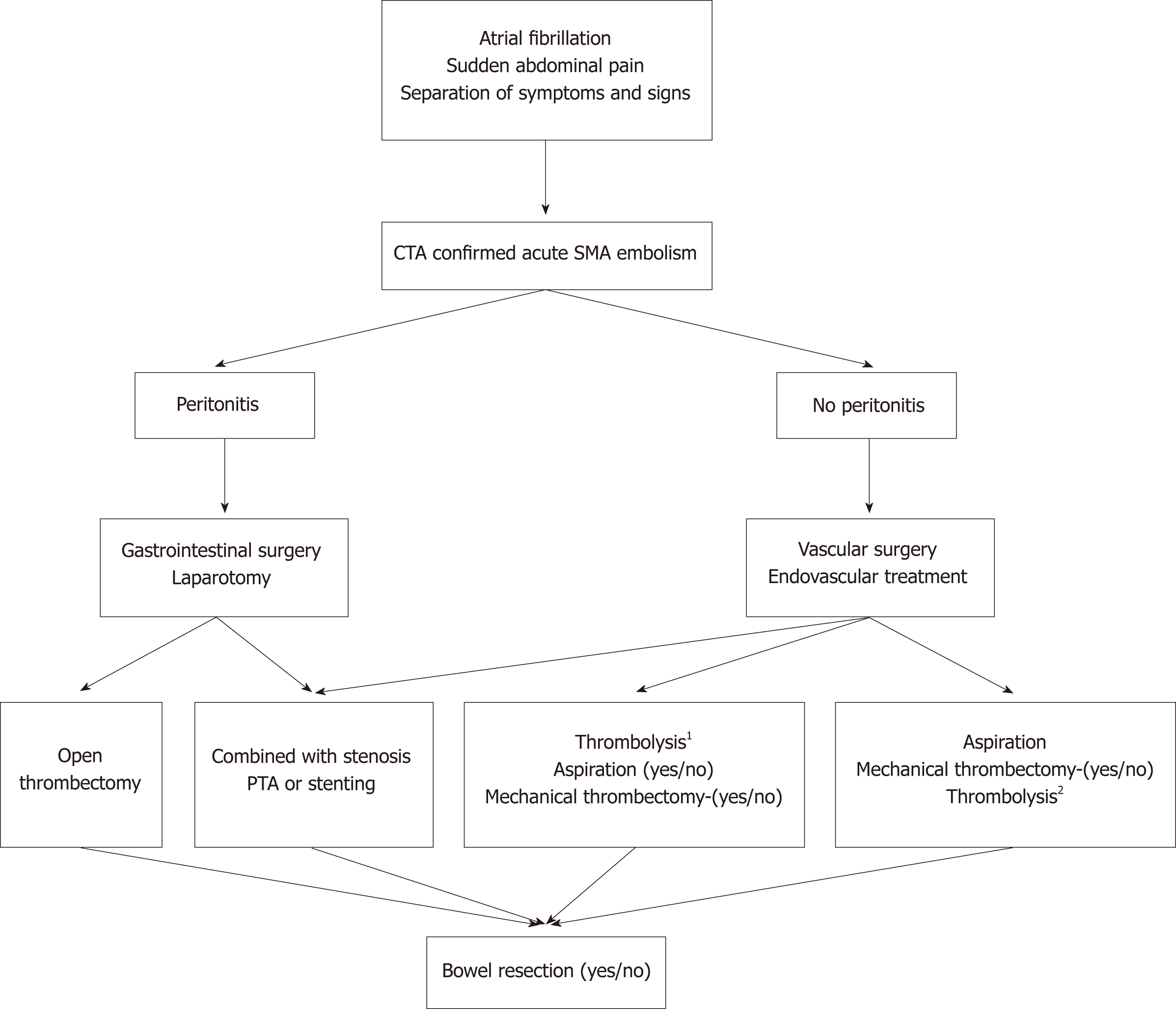
Figure 2 The processing flow of acute embolic occlusion of the superior mesenteric artery.
1Patients with poor general condition first underwent thrombolysis; 2Residual thrombus was treated by thrombolysis during the operation or catheter-directed thrombolysis after aspiration. PTA: Percutaneous transluminal angioplasty; CTA: Computed tomography angiography; SMA: Superior mesenteric artery.
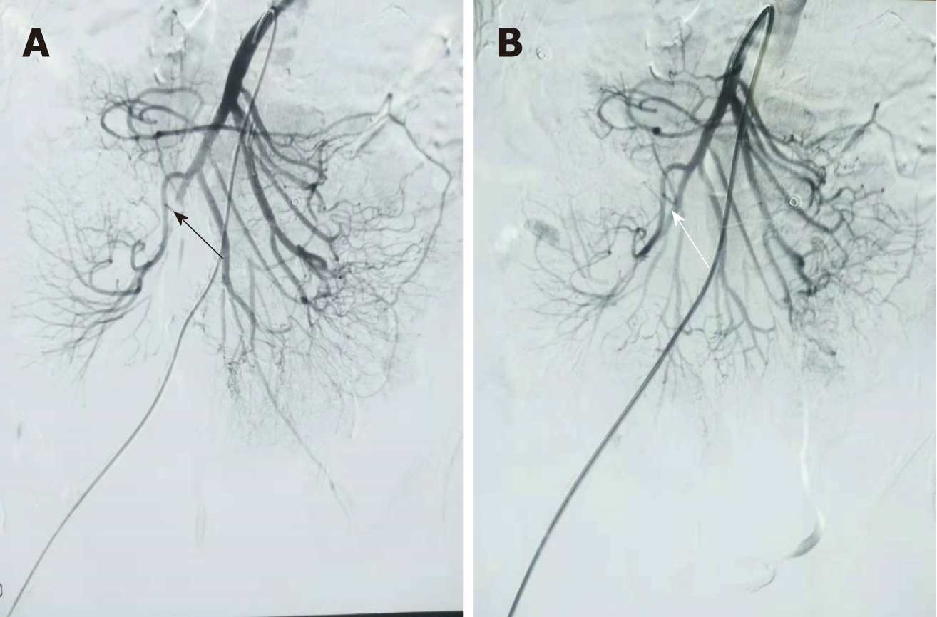
Figure 3 Digital subtraction angiography images.
A: Filling defect of the superior mesenteric artery (SMA) indicated by the black arrow; B: Complete patency of the SMA indicated by the white arrow.
Figure 4 Embolus images.
A: Aspirated embolus; B: Pathology consisting of white and red clots.
- Citation: Liu YR, Tong Z, Hou CB, Cui SJ, Guo LR, Qi YX, Qi LX, Guo JM, Gu YQ. Aspiration therapy for acute embolic occlusion of the superior mesenteric artery. World J Gastroenterol 2019; 25(7): 848-858
- URL: https://www.wjgnet.com/1007-9327/full/v25/i7/848.htm
- DOI: https://dx.doi.org/10.3748/wjg.v25.i7.848


