Copyright
©The Author(s) 2019.
World J Gastroenterol. Feb 14, 2019; 25(6): 696-706
Published online Feb 14, 2019. doi: 10.3748/wjg.v25.i6.696
Published online Feb 14, 2019. doi: 10.3748/wjg.v25.i6.696
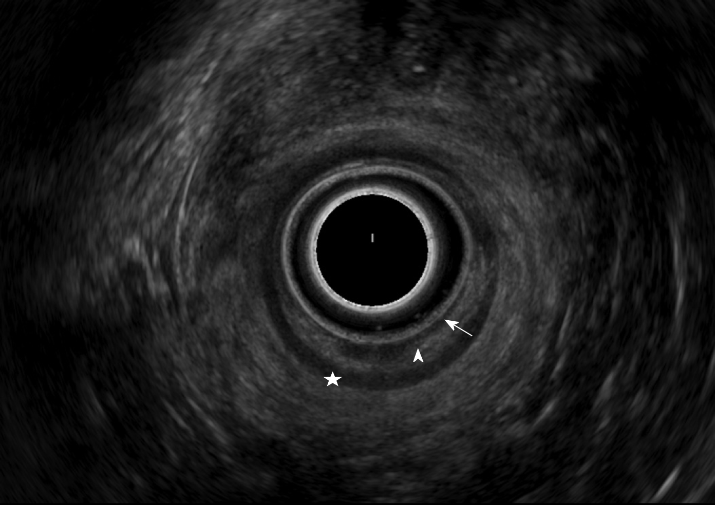
Figure 1 Normal view of the rectal wall with a radial probe in Rectosigmoid Endoscopic Ultrasonography.
Arrow: Mucosae; Arrowhead: Muscular mucosa; Star: Submucosa; Disc: Muscular layer; Device: PENTAX EG-3670 URK ultrasound video-endoscope 7.5 MHz.
Figure 2 View with a radial probe of an endometriotic nodule in rectosigmoid endoscopic ultrasonography.
This nodule is located in the front side of the upper rectum next to the torus and exhibits infiltration of the submucosa. Width: 11 mm; Thickness: 8.6 mm. Arrow: Mucosae; Star: Submucosa; Arrowhead: Muscular layer; Device: PENTAX EG-3670 URK ultrasound video-endoscope 7.5 MHz.
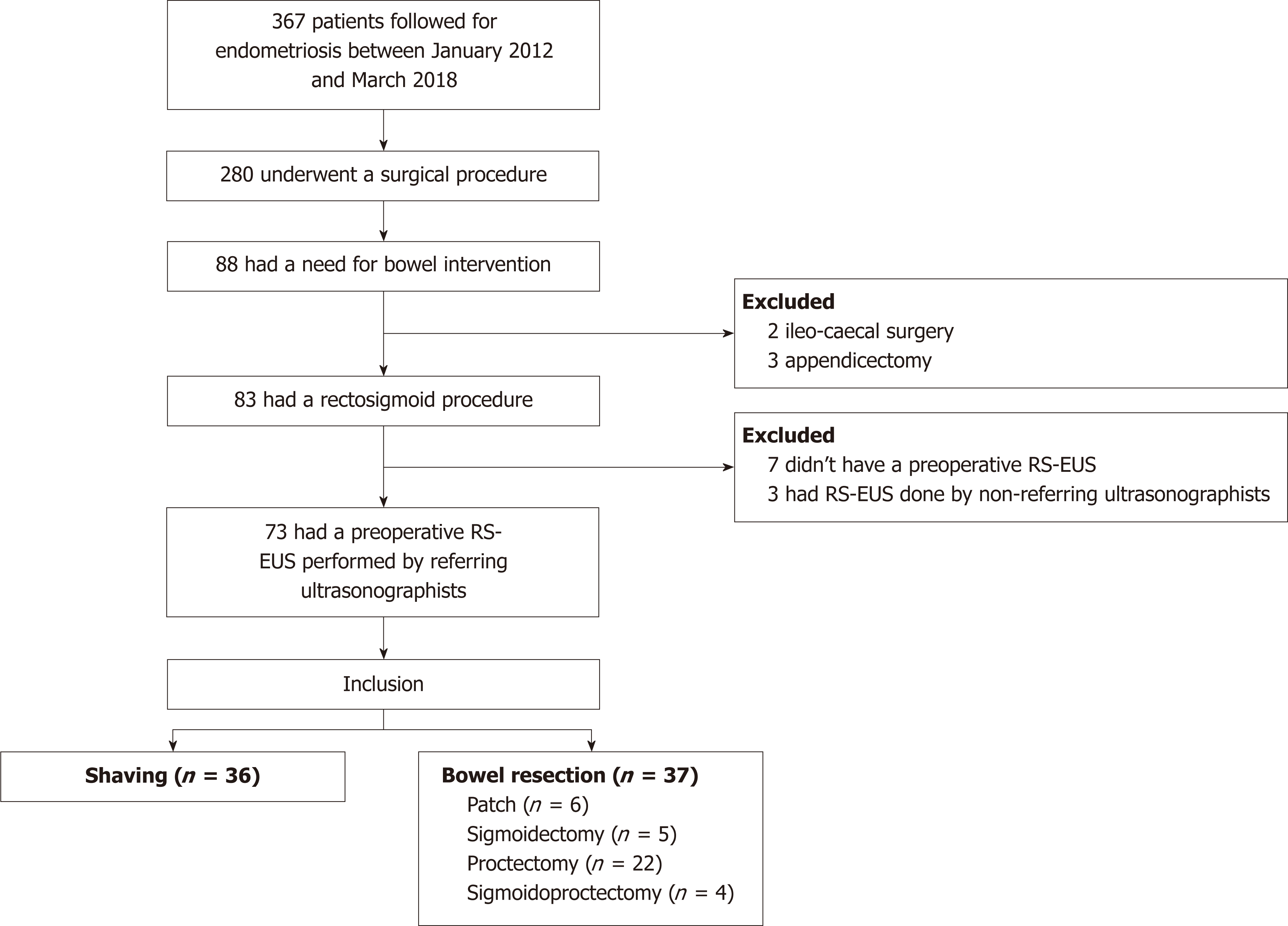
Figure 3 Flow chart.
RS-EUS: Rectosigmoid endoscopic ultrasonography.
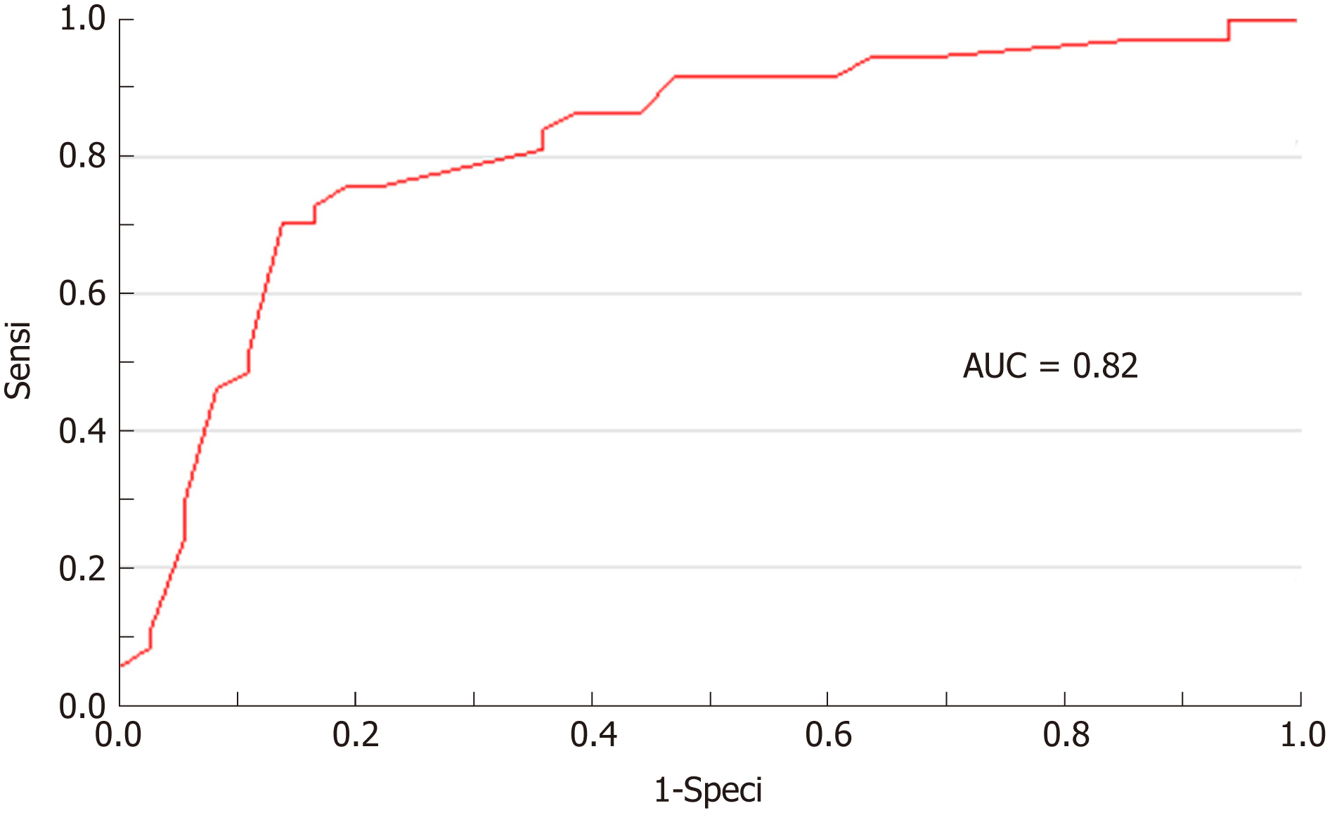
Figure 4 Receiver operating characteristic curve of nodule thickness.
AUC: Area under curve.
- Citation: Desplats V, Vitte RL, du Cheyron J, Roseau G, Fauconnier A, Moryoussef F. Preoperative rectosigmoid endoscopic ultrasonography predicts the need for bowel resection in endometriosis. World J Gastroenterol 2019; 25(6): 696-706
- URL: https://www.wjgnet.com/1007-9327/full/v25/i6/696.htm
- DOI: https://dx.doi.org/10.3748/wjg.v25.i6.696


