Copyright
©The Author(s) 2019.
World J Gastroenterol. Feb 14, 2019; 25(6): 696-706
Published online Feb 14, 2019. doi: 10.3748/wjg.v25.i6.696
Published online Feb 14, 2019. doi: 10.3748/wjg.v25.i6.696
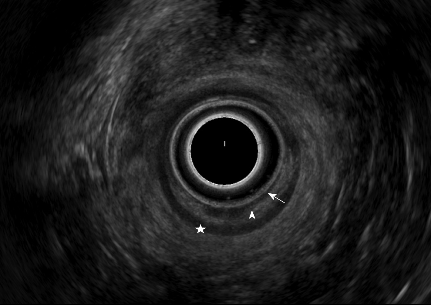
Figure 1 Normal view of the rectal wall with a radial probe in Rectosigmoid Endoscopic Ultrasonography.
Arrow: Mucosae; Arrowhead: Muscular mucosa; Star: Submucosa; Disc: Muscular layer; Device: PENTAX EG-3670 URK ultrasound video-endoscope 7.5 MHz.
- Citation: Desplats V, Vitte RL, du Cheyron J, Roseau G, Fauconnier A, Moryoussef F. Preoperative rectosigmoid endoscopic ultrasonography predicts the need for bowel resection in endometriosis. World J Gastroenterol 2019; 25(6): 696-706
- URL: https://www.wjgnet.com/1007-9327/full/v25/i6/696.htm
- DOI: https://dx.doi.org/10.3748/wjg.v25.i6.696


