Copyright
©The Author(s) 2019.
World J Gastroenterol. Dec 28, 2019; 25(48): 6949-6958
Published online Dec 28, 2019. doi: 10.3748/wjg.v25.i48.6949
Published online Dec 28, 2019. doi: 10.3748/wjg.v25.i48.6949
Figure 1 Computed tomographic scans of hepatocellular carcinoma.
A: Computed tomographic scans of hepatocellular carcinoma (HCC) in the liver; B: Computed tomographic scans of HCC in the metastases to sacral bone; C: Computed tomographic scans of HCC in the lung. White arrowheads indicate the tumor.
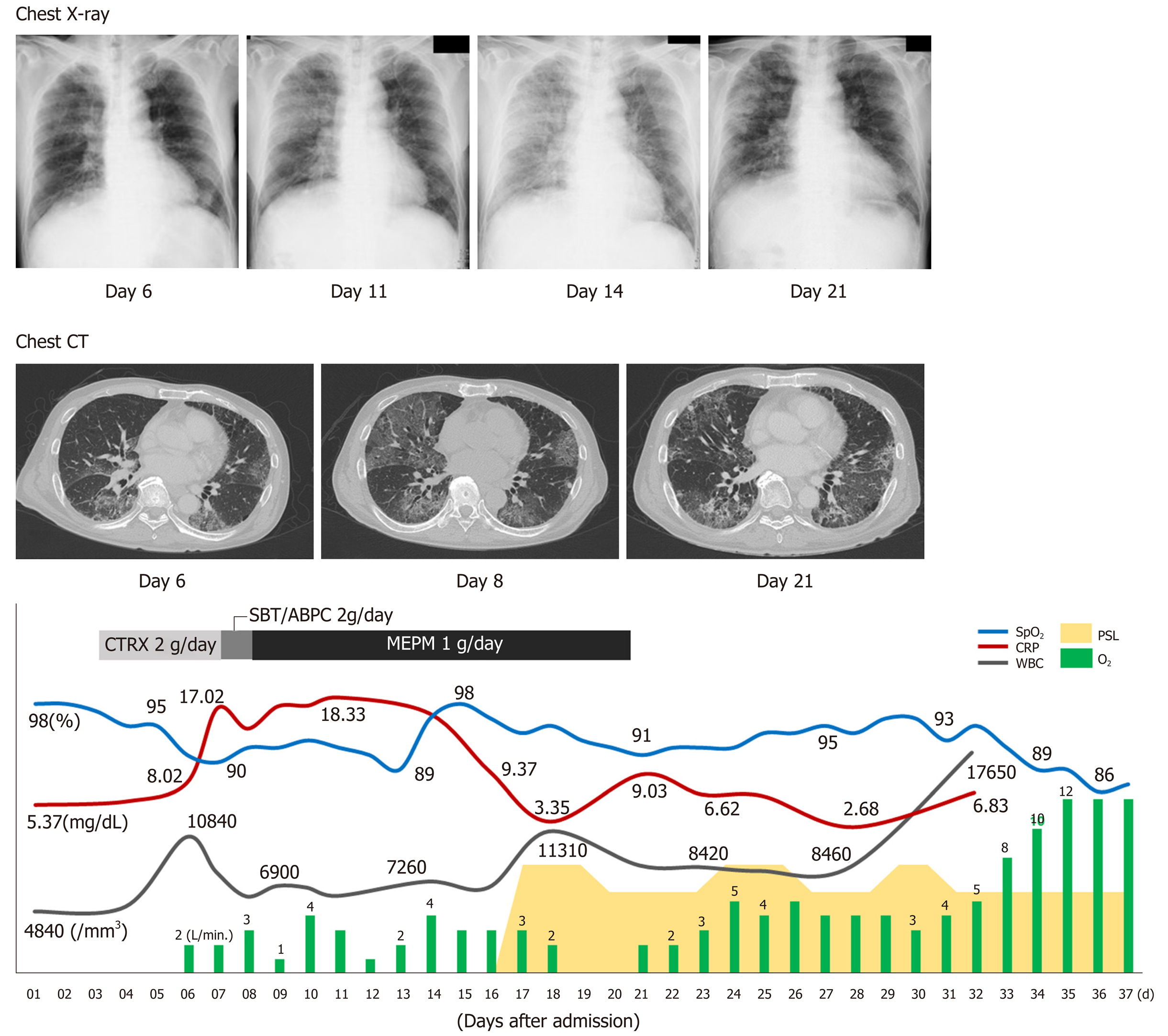
Figure 2 Clinical courses of physical and laboratory findings, chest radiograph, and computed tomographic scans.
CT: Computed tomographic; BT: Body temperature; CRP: C-reactive protein; CTRX: Ceftriaxone sodium hydrate; MEPM: Meropenem; PSL: Prednisolone; SBT/ABPC: Sulbactam/ampicillin; SpO2: Oxygen saturation; WBC: White blood cell count.
Figure 3 Histological analyses.
A: Macroscopic findings of the lung; B: Hematoxylin and eosin staining of the tumor; C, D: Diffuse alveolar damages with multiple pulmonary artery tumor emboli. White arrows in cindicate the alveolar damage and arrowheads in Cindicate tumor cells. Black arrow in D indicates the recanalization and a white arrowhead indicates the fibrocellular intimal proliferation; E: Medial thickening of arterioles. A white arrowhead indicates the thickening; F: Tumor emboli (hematoxylin and eosin staining) were accompanied by the CD31-positive endothelial cell growth; G: CD31 staining, a white arrow head and fibrocellular intimal proliferation; H: Elastica van Gieson staining, a white arrow head.
- Citation: Morita S, Kamimura K, Abe H, Watanabe-Mori Y, Oda C, Kobayashi T, Arao Y, Tani Y, Ohashi R, Ajioka Y, Terai S. Pulmonary tumor thrombotic microangiopathy of hepatocellular carcinoma: A case report and review of literature. World J Gastroenterol 2019; 25(48): 6949-6958
- URL: https://www.wjgnet.com/1007-9327/full/v25/i48/6949.htm
- DOI: https://dx.doi.org/10.3748/wjg.v25.i48.6949


