Copyright
©The Author(s) 2019.
World J Gastroenterol. Nov 28, 2019; 25(44): 6527-6540
Published online Nov 28, 2019. doi: 10.3748/wjg.v25.i44.6527
Published online Nov 28, 2019. doi: 10.3748/wjg.v25.i44.6527
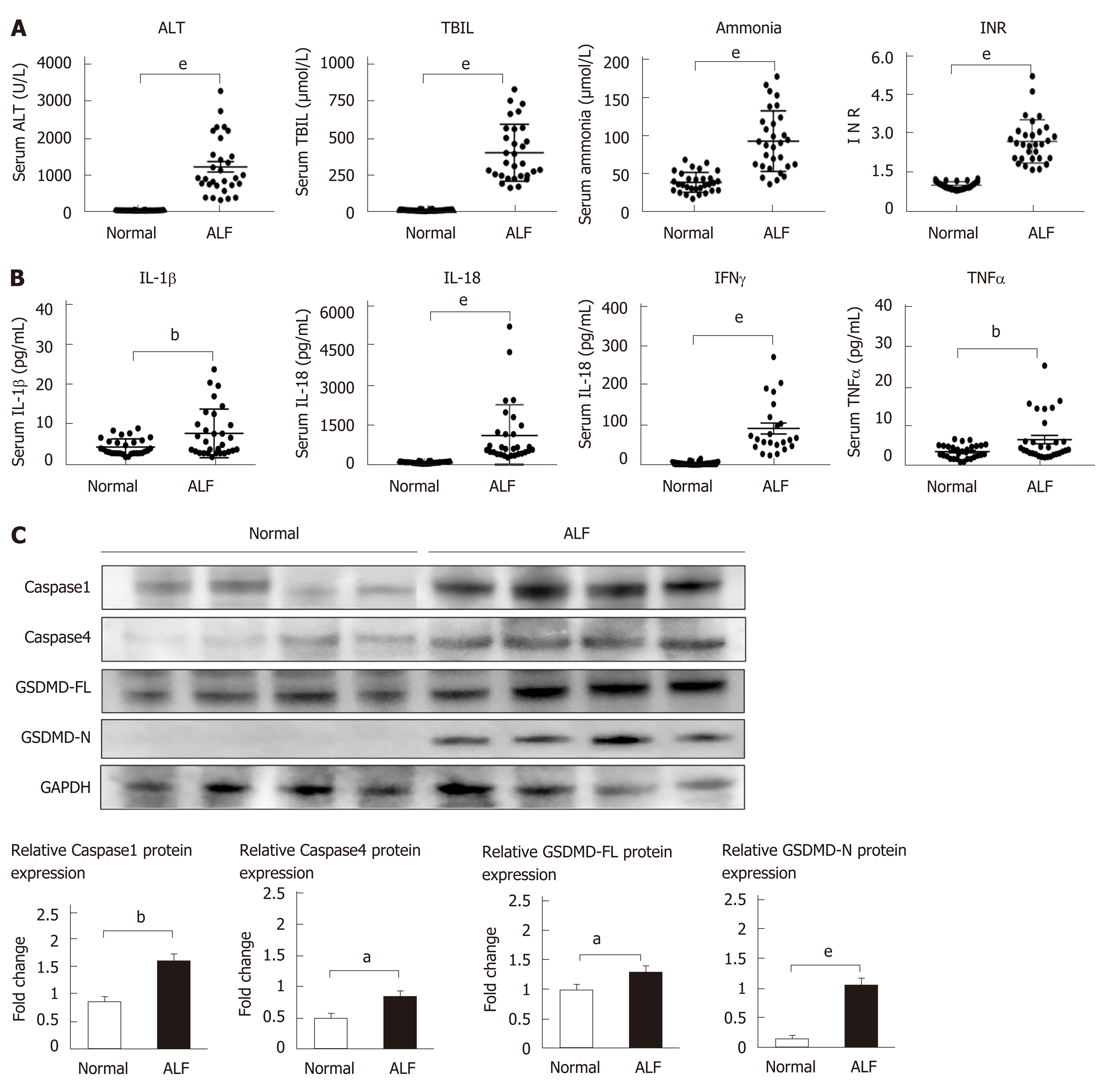
Figure 1 The levels of pyroptosis-associated inflammatory cytokines and proteins increase in human acute liver failure.
A: Serum levels of alanine aminotransferase, total bilirubin, ammonia, and international normalized ratio; B: Serum levels of inflammatory cytokines interleukin (IL)-1β, IL-18, tumor necrosis factor-alpha, and interferon-γ detected by flow cytometry; C: Expression levels of caspase 1, caspase 4, full-length gasdermin D, and cleaved N-terminal fragment of gasdermin D in liver tissues from acute liver failure cases and controls detected using Western blot with GAPDH as the internal reference. Data are presented as the mean ± square error. aP < 0.05, bP < 0.01, and eP < 0.001. ALT: Alanine aminotransferase; TBIL: Total bilirubin; INR: International normalized ratio; IL-1β: Interleukin-1β; IL-18: Interleukin-18; TNFα: Tumor necrosis factor-alpha; IFN-γ: Interferon-γ; GSDMD-FL: Full-length gasdermin D; GSDMD-N: Cleaved N-terminal fragment of gasdermin D; ALF: Acute liver failure.
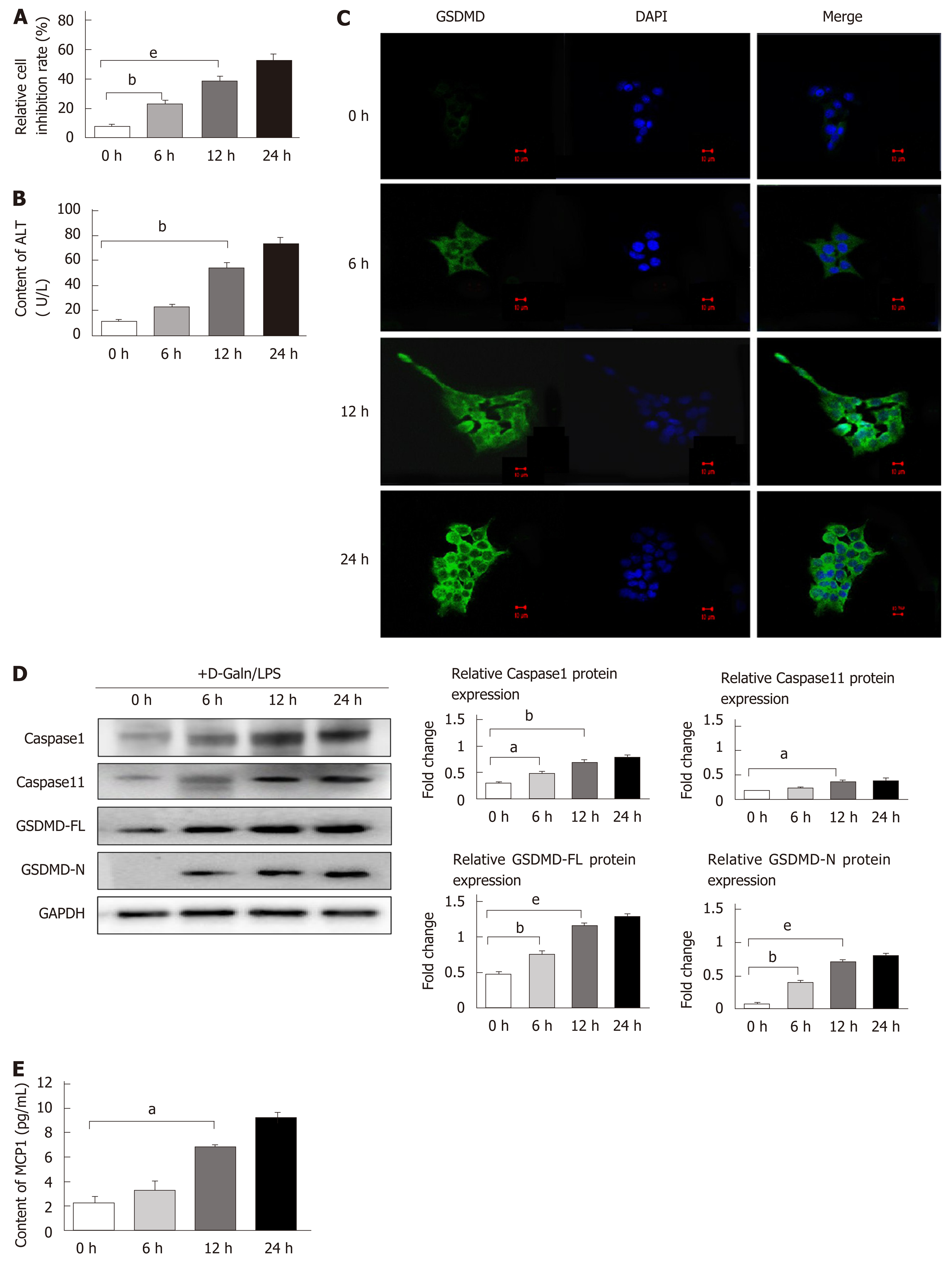
Figure 2 Expression of pyroptosis pathway-associated proteins increases in a D-galactose/lipopolysaccharide-induced AML12 hepatocyte injury model.
Intervention was performed using D-Galn (15 mmol/L) + LPS (100 µg/mL) for 0, 6, 12, and 24 h. A: Changes in the cell inhibition rate at 0, 6, 12, and 24 h evaluated by CCK8; B: Content of ALT in the supernatant; C: Expression of gasdermin D (GSDMD) protein detected under a confocal fluorescence microscope. Scale bars = 10 µm; D: Caspase 1/11, GSDMD-FL, and GSDMD-N protein expression evaluated by Western blot with GAPDH as the internal reference; E: Content of MCP1 in the cell culture supernatant. Data are presented as the mean ± square error. aP < 0.05, bP < 0.01, and eP < 0.001. ALT: Alanine aminotransferase; D-Galn/LPS: D-galactose/lipopolysaccharide; GSDMD-FL: Full-length GSDMD; GSDMD-N: Cleaved N-terminal fragment of GSDMD; MCP1: Monocyte chemotactic protein 1; DAPI: 4’,6-diamidino-2-phenylindole.
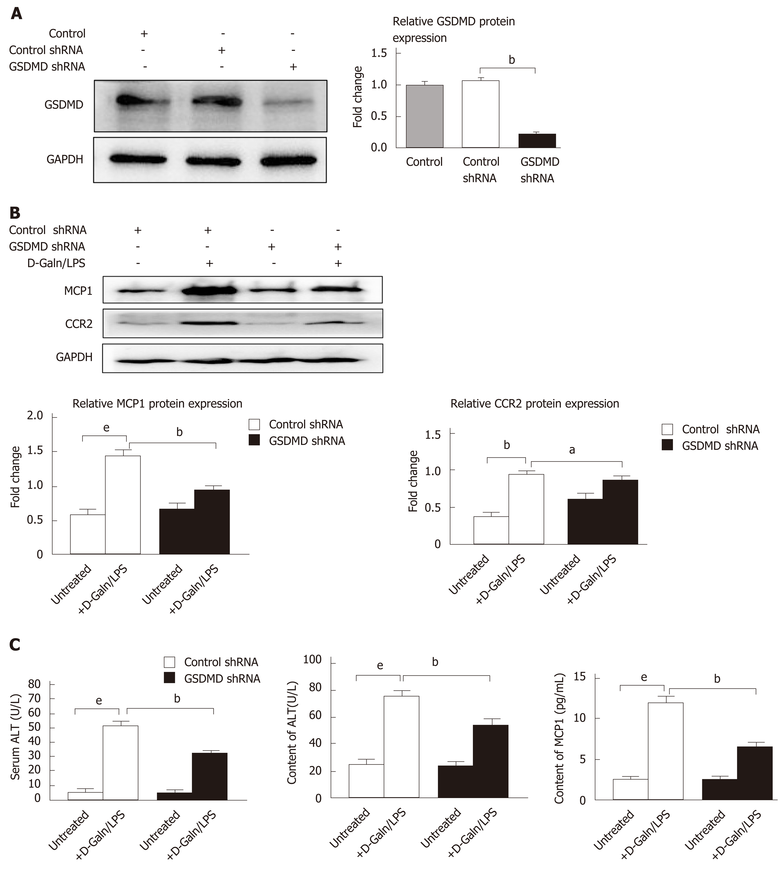
Figure 3 Gasdermin D knockdown by shRNA decreases the expression of monocyte chemotactic protein 1 and CC chemokine receptor-2 in a D-galactose/lipopolysaccharide-induced AML12 hepatocyte injury model.
Control shRNA and gasdermin D (GSDMD) shRNA were transfected into AML12 using Lipofectamine 3000, and the intervention was performed using D-galactose (15 mmol/L)/lipopolysaccharide (100 µg/L) for 24 h. A: Expression changes of GSDMD protein evaluated by Western blot; B: Protein expression levels of monocyte chemotactic protein 1 (MCP1) and CC chemokine receptor-2 evaluated by Western blot; C: Cell inhibition rate evaluated by CCK8 assay; D: Contents of alanine aminotransferase and MCP1 in the cell supernatant. Data are presented as the mean ± square error. aP < 0.05, bP < 0.01, and eP < 0.001. D-Galn/LPS: D-galactose/lipopolysaccharide; GSDMD: Gasdermin D; ALT: Alanine aminotransferase; MCP1: Monocyte chemotactic protein 1; CCR2: CC chemokine receptor-2.
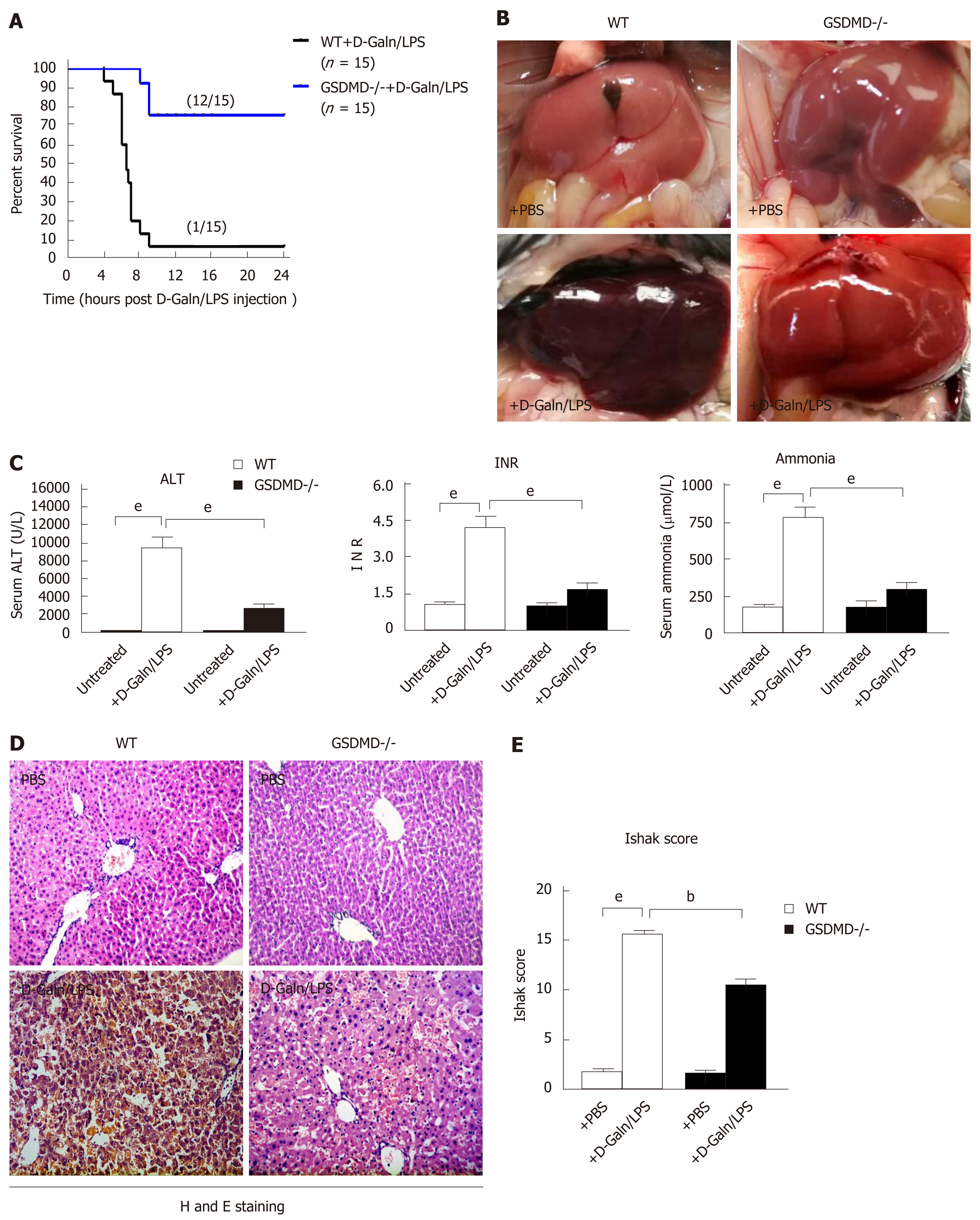
Figure 4 Gasdermin D knockout dramatically eliminates inflammatory damage in the liver and improves survival in D-galactose/lipopolysaccharide-induced acute liver failure mice.
A: Survival curve; B: Changes in liver tissue appearance; C: Alanine aminotransferase, international normalized ratio, and blood ammonia in mice; D: Pathological examination of the liver by haematoxylin and eosin staining. Scale bars = 100 µm; E: Ishak score of the liver tissue pathology. Data are presented as the mean ± square error. aP < 0.05, bP < 0.01, and eP < 0.001. WT: Wild type; GSDMD-/-: Gasdermin D genetic knockout; D-Galn/LPS: D-galactose/lipopolysaccharide; PBS: Phosphate buffer saline; ALT: Alanine aminotransferase; INR: International normalized ratio; H and E: Haematoxylin and eosin.
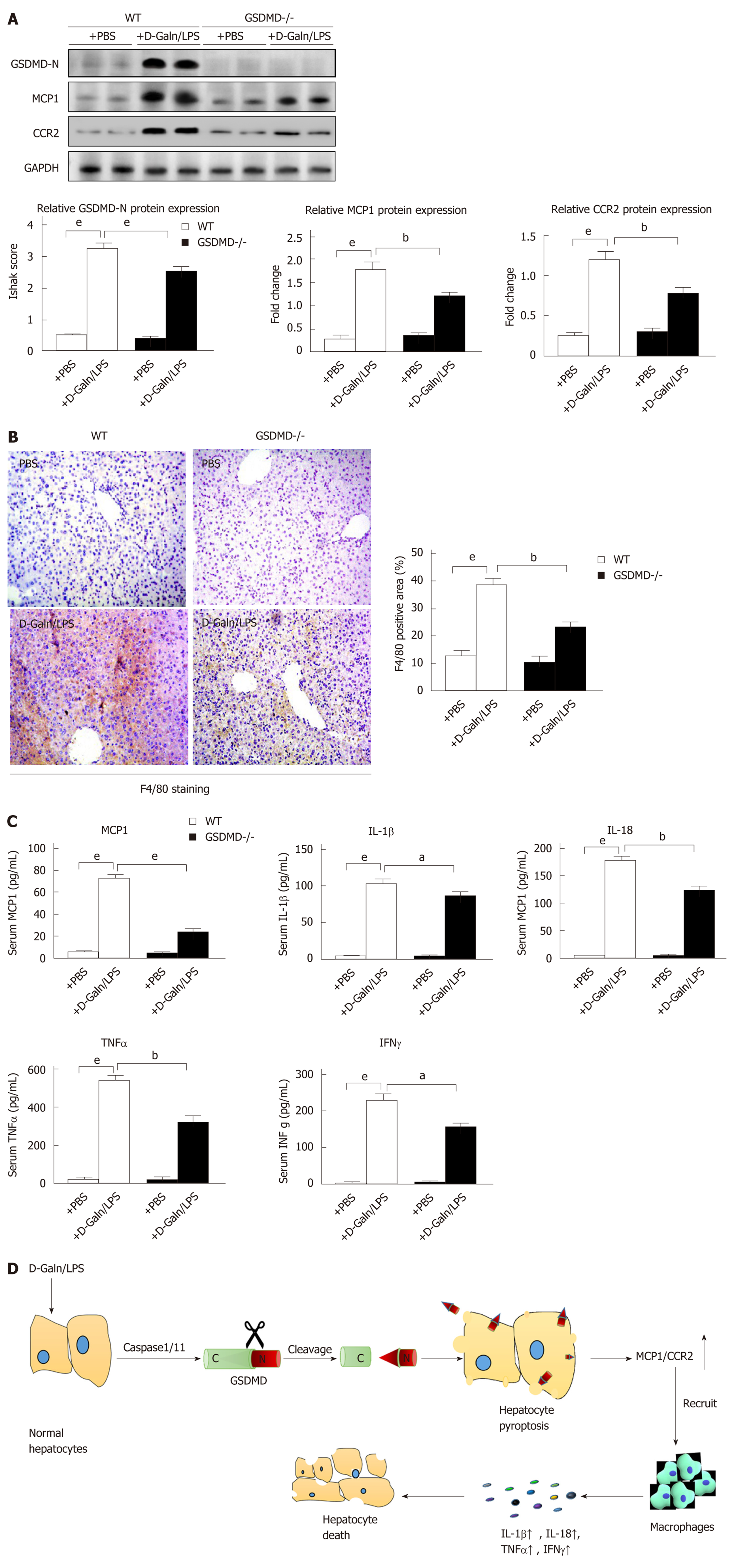
Figure 5 Gasdermin D knockout significantly decreases the expression of monocyte chemotactic protein 1/ CC chemokine receptor-2 and macrophage infiltration in D-galactose/lipopolysaccharide-induced acute liver failure mice.
A: Protein expression levels of cleaved N-terminal fragment of gasdermin D, monocyte chemotactic protein 1 (MCP1), and CC chemokine receptor-2 in the liver evaluated by Western blot; B: Immunohistochemical staining for macrophage-specific F4/80 in the liver. Scale bars = 100 µm; C: Detection of serum MCP1, interleukin (IL)-1β, IL-18, tumor necrosis factor-alpha, and interferon-γ by enzyme-linked immunosorbent assay; D: The diagram showing the mechanism of hepatocyte pyroptosis for expanding the inflammatory responses; Data are presented as the mean ± square error. aP < 0.05, bP < 0.01, and eP < 0.001. WT: Wild type; GSDMD-/-: GSDMD genetic knockout; D-Galn/LPS: D-galactose/lipopolysaccharide; PBS: Phosphate buffer saline; GSDMD-N: Cleaved N-terminal fragment of GSDMD; MCP1: Monocyte chemotactic protein 1; CCR2: CC chemokine receptor-2; IL-1β: Interleukin-1β; IL-18: Interleukin-18; TNFα: Tumor necrosis factor-alpha; IFNγ: Interferon-γ.
- Citation: Li H, Zhao XK, Cheng YJ, Zhang Q, Wu J, Lu S, Zhang W, Liu Y, Zhou MY, Wang Y, Yang J, Cheng ML. Gasdermin D-mediated hepatocyte pyroptosis expands inflammatory responses that aggravate acute liver failure by upregulating monocyte chemotactic protein 1/CC chemokine receptor-2 to recruit macrophages. World J Gastroenterol 2019; 25(44): 6527-6540
- URL: https://www.wjgnet.com/1007-9327/full/v25/i44/6527.htm
- DOI: https://dx.doi.org/10.3748/wjg.v25.i44.6527


