Copyright
©The Author(s) 2019.
World J Gastroenterol. Nov 21, 2019; 25(43): 6430-6439
Published online Nov 21, 2019. doi: 10.3748/wjg.v25.i43.6430
Published online Nov 21, 2019. doi: 10.3748/wjg.v25.i43.6430
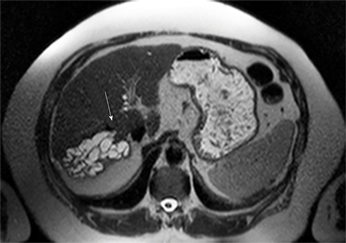
Figure 1 Transversal T2w magnetic resonance imaging of the liver of patient 2 who presented with segmental intrahepatic cholestasis caused by intrahepatic bile duct compression after transjugular intrahepatic portosystemic shunt.
The arrow indicates the intrahepatic portion of the stent. Note the tubular structure with fluid typical high T2w signal converging in direct proximity of the transjugular intrahepatic portosystemic shunt which represents the congested, intrahepatic bile ducts of liver segment VII. The segments’ liver parenchyma is completely extinct by the dilated ducts. Other causes for bile duct obstruction were ruled out by T1w with liver specific contrast.
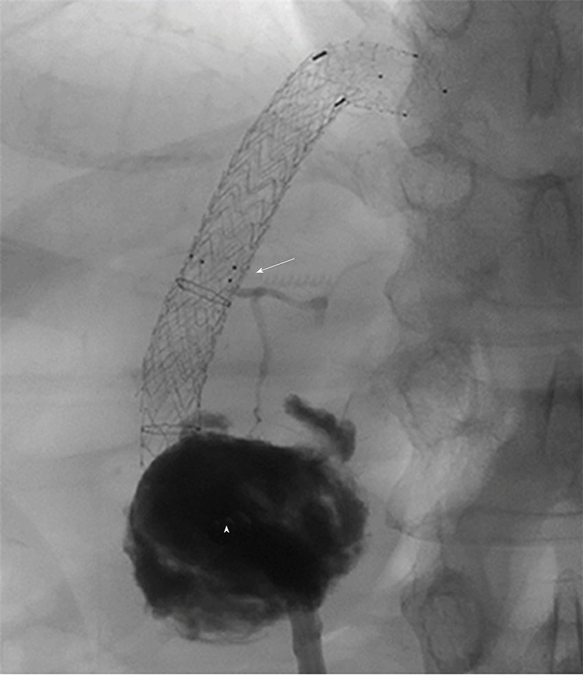
Figure 2 Angiography after contrast-injection through the interventional drain in patient 4.
The abscess (triangle) is filled with contrast agent. The abscess is connected with the segmental bile duct (segment I) that is interrupted by the transjugular intrahepatic portosystemic shunt-stent as indicated by the arrowhead.
- Citation: Bucher JN, Hollenbach M, Strocka S, Gaebelein G, Moche M, Kaiser T, Bartels M, Hoffmeister A. Segmental intrahepatic cholestasis as a technical complication of the transjugular intrahepatic porto-systemic shunt. World J Gastroenterol 2019; 25(43): 6430-6439
- URL: https://www.wjgnet.com/1007-9327/full/v25/i43/6430.htm
- DOI: https://dx.doi.org/10.3748/wjg.v25.i43.6430


