
















©The Author(s) 2019.
World J Gastroenterol. Nov 21, 2019; 25(43): 6430-6439
Published online Nov 21, 2019. doi: 10.3748/wjg.v25.i43.6430
Published online Nov 21, 2019. doi: 10.3748/wjg.v25.i43.6430
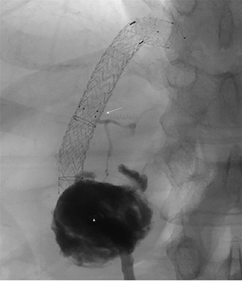
Figure 2 Angiography after contrast-injection through the interventional drain in patient 4.
The abscess (triangle) is filled with contrast agent. The abscess is connected with the segmental bile duct (segment I) that is interrupted by the transjugular intrahepatic portosystemic shunt-stent as indicated by the arrowhead.
- Citation: Bucher JN, Hollenbach M, Strocka S, Gaebelein G, Moche M, Kaiser T, Bartels M, Hoffmeister A. Segmental intrahepatic cholestasis as a technical complication of the transjugular intrahepatic porto-systemic shunt. World J Gastroenterol 2019; 25(43): 6430-6439
- URL: https://www.wjgnet.com/1007-9327/full/v25/i43/6430.htm
- DOI: https://dx.doi.org/10.3748/wjg.v25.i43.6430