Copyright
©The Author(s) 2019.
World J Gastroenterol. Nov 7, 2019; 25(41): 6222-6237
Published online Nov 7, 2019. doi: 10.3748/wjg.v25.i41.6222
Published online Nov 7, 2019. doi: 10.3748/wjg.v25.i41.6222
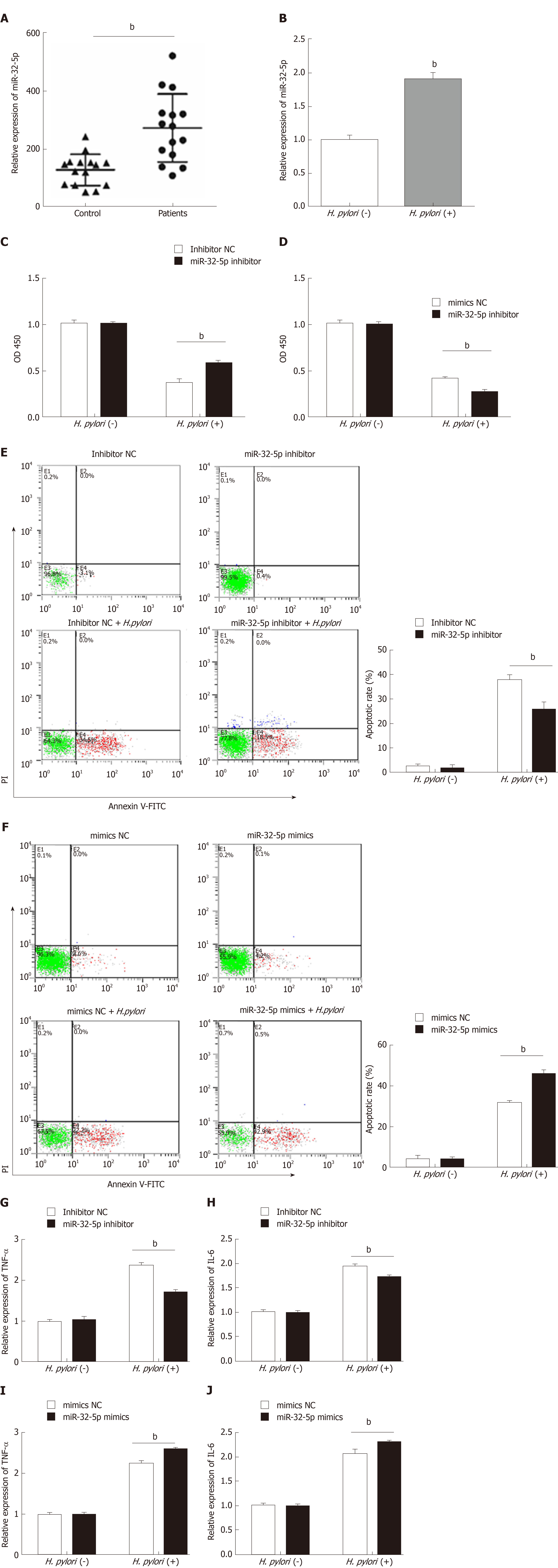
Figure 1 Aberrant expression of miR-32-5p in enteritis regulates biological function of intestinal epithelial cells.
A: Expression of miR-32-5p in serum of pediatric enteritis; B: Expression of miR-32-5p in intestinal epithelial cells infected by Helicobacter pylori (H. pylori); C: Cell viability measurement in intestinal epithelial cells transfected with miR-32-5p inhibitor in the presence or absence of H. pylori; D: Viability measurement in intestinal epithelial cells transfected with miR-32-5p mimic in the presence or absence of H. pylori; E: Apoptosis of intestinal epithelial cells with miR-32-5p inhibitor transfection in the presence or absence of H. pylori; F: Apoptosis of intestinal epithelial cells with miR-32-5p mimic transfection in the presence or absence of H. pylori; G: The mRNA level of TNF-α in H. pylori-infected intestinal epithelial cells in the presence of miR-32-5p inhibitor; I: The mRNA level of TNF-α in H. pylori-infected intestinal epithelial cells in the presence of miR-32-5p mimic; H: The mRNA level of IL-6 in H. pylori-infected intestinal epithelial cells in the presence of miR-32-5p inhibitor; J: The mRNA level of IL-6 in H. pylori-infected intestinal epithelial cells in the presence of miR-32-5p mimic. bP < 0.01. H. pylori: Helicobacter pylori.
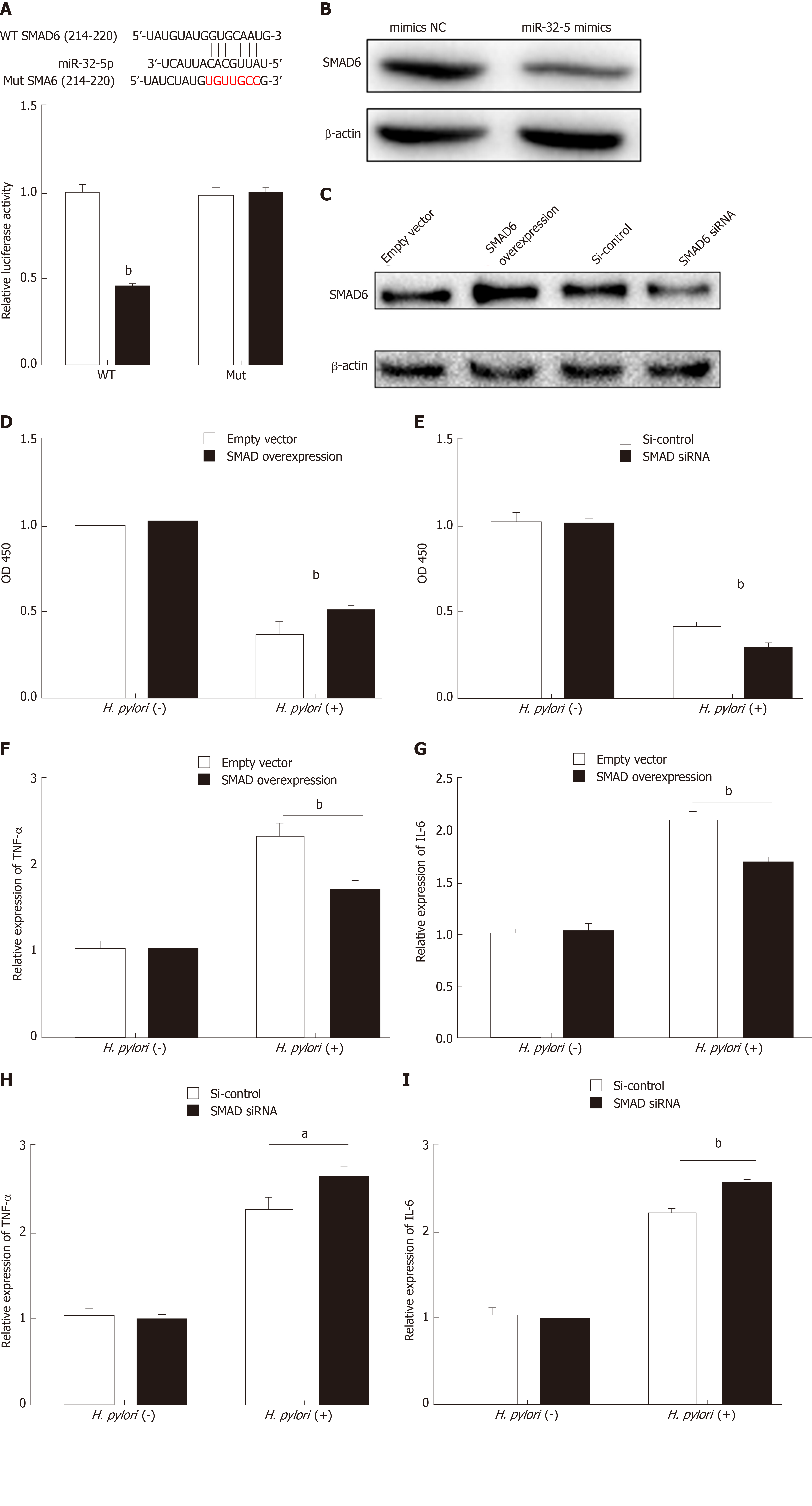
Figure 2 SMAD family member 6 is sponged by miR-32-5p in intestinal epithelial cells.
A: Luciferase assay for determining the binding between miR-32-5p and SMAD family member 6 (SMAD6); B: Protein level of SMAD6 in intestinal epithelial cells transfected with miR-32-5p mimic; C: Expression of SMAD6 in intestinal epithelial cells after SMAD6 overexpression and knockdown; D: Cell viability measurement in intestinal epithelial cells after SMAD6 overexpression in the presence or absence of H. pylori; E: Cell viability measurement in intestinal epithelial cells after SMAD6 knockdown in the presence or absence of Helicobacter pylori (H. pylori); F: The mRNA level of TNF-α in H. pylori-infected intestinal epithelial cells after SMAD6 overexpression; H: The mRNA level of TNF-α in H. pylori-infected intestinal epithelial cells after SMAD6 knockdown; G: IL-6 expression in H. pylori-infected intestinal epithelial cells after SMAD6 overexpression; I: IL-6 expression in H. pylori-infected intestinal epithelial cells after SMAD6 knockdown. aP < 0.05, bP < 0.01. SMAD6: SMAD family member 6; H. pylori: Helicobacter pylori.
Figure 3 Transforming growth factor-β1/p38 participates in apoptosis of intestinal epithelial cells infected by Helicobacter pylori.
A: Apoptosis detection in transforming growth factor-β1 (TGF-β1)-treated intestinal epithelial cells in the presence of transforming growth factor-β-activated kinase 1 (TAK1) inhibitor and p38 inhibitor; B: Protein levels of total TAK1, p38, phosphorylated TAK1, and phosphorylated p38 in TGF-β1-treated intestinal epithelial cells in the presence of TAK1 inhibitor and p38 inhibitor; C: Cell viability measurement in Helicobacter pylori (H. pylori)-infected intestinal epithelial cells with TAK1 inhibitor and p38 inhibitor treatment followed by transfection with miR-32-5p mimic; D: Cell viability measurement in H. pylori-infected intestinal epithelial cells with TAK1 inhibitor and p38 inhibitor treatment followed by transfection with miR-32-5p inhibitor; E: Apoptosis evaluation in H. pylori-infected intestinal epithelial cells with TAK1 inhibitor and p38 inhibitor treatment followed by transfection with miR-32-5p mimic; F: Apoptosis evaluation in H. pylori-infected intestinal epithelial cells with TAK1 inhibitor and p38 inhibitor treatment followed by transfection with miR-32-5p inhibitor. bP < 0.01, dP < 0.01, fP < 0.01, gP < 0.05, hP < 0.01. TGF-β1: Transforming growth factor-β1; TAK1: Transforming growth factor-β-activated kinase 1; H. pylori: Helicobacter pylori.
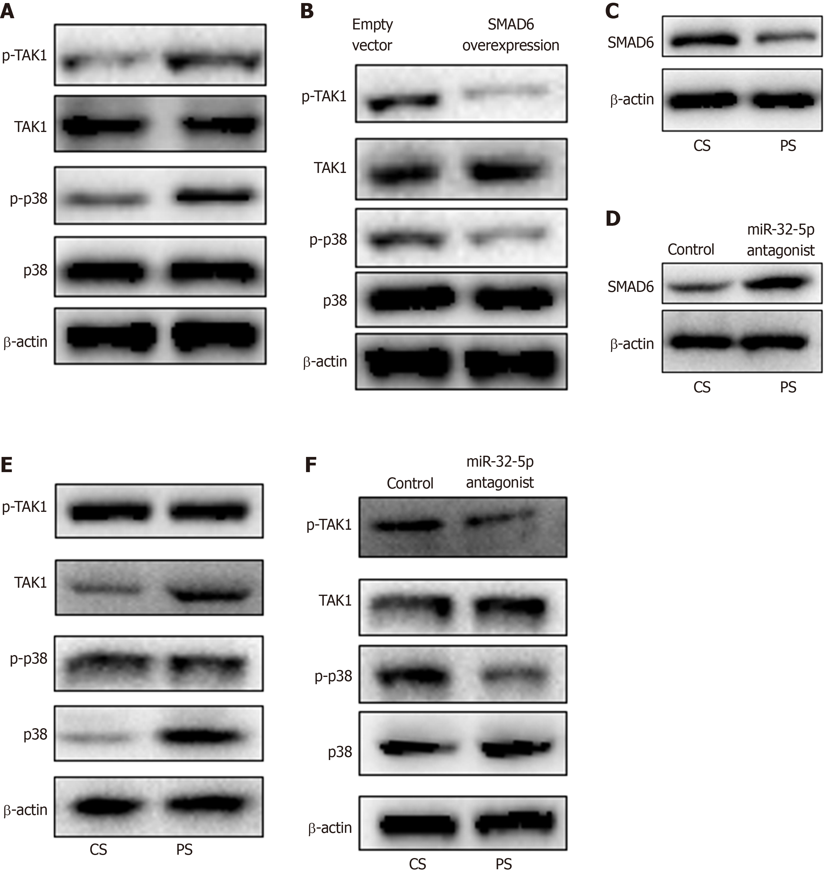
Figure 4 MiR-32-5p/SMAD family member 6 is involved in transforming growth factor-β-activated kinase 1-p38 pathway in intestinal epithelial cells.
A: Detection of transforming growth factor-β-activated kinase 1 (TAK1)-p38 activation in Helicobacter pylori (H. pylori)-treated intestinal epithelial cells by Western blot; B: Inhibition of TAK1-p38 activation in H. pylori-treated intestinal epithelial cells with SMAD6 overexpression as revealed by Western blot; C: SMAD6 expression in intestinal epithelial cells treated with control serum and patient serum (PS); D: SMAD6 expression in PS-treated intestinal epithelial cells with miR-32-5p antagonist transfection; E: TAK1-p38 activation in PS-treated intestinal epithelial cells as revealed by Western blot; F: Inhibition of TAK1-p38 activation in PS-treated intestinal epithelial cells with miR-32-5p antagonist transfection as revealed by Western blot. CS: Control serum; PS: Patient serum; SMAD6: SMAD family member 6; TAK1: Transforming growth factor-β-activated kinase 1; H. pylori: Helicobacter pylori.
- Citation: Feng J, Guo J, Wang JP, Chai BF. MiR-32-5p aggravates intestinal epithelial cell injury in pediatric enteritis induced by Helicobacter pylori. World J Gastroenterol 2019; 25(41): 6222-6237
- URL: https://www.wjgnet.com/1007-9327/full/v25/i41/6222.htm
- DOI: https://dx.doi.org/10.3748/wjg.v25.i41.6222


