Copyright
©The Author(s) 2019.
World J Gastroenterol. Jun 7, 2019; 25(21): 2549-2564
Published online Jun 7, 2019. doi: 10.3748/wjg.v25.i21.2549
Published online Jun 7, 2019. doi: 10.3748/wjg.v25.i21.2549
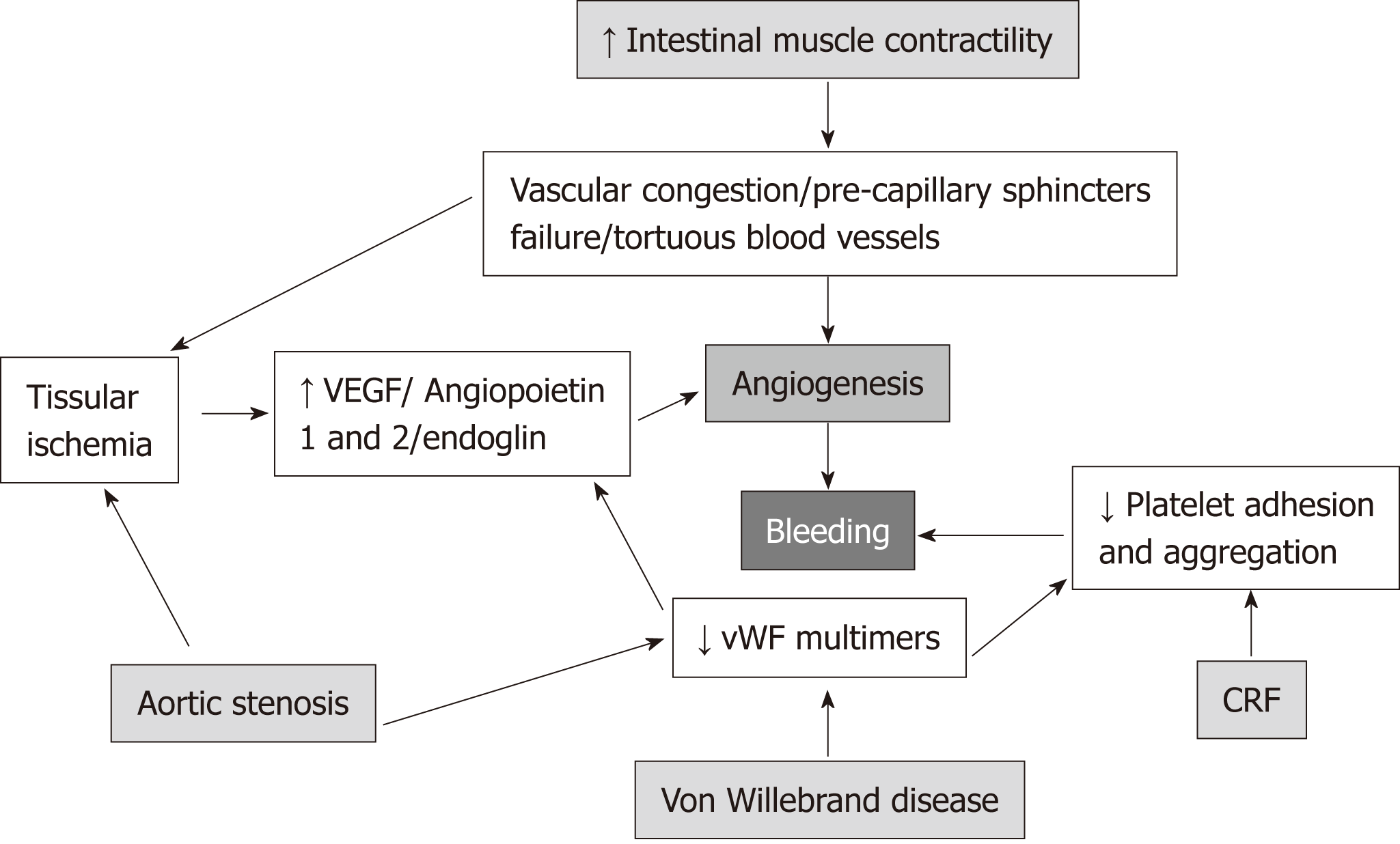
Figure 1 Pathogenesis of gastrointestinal angiodysplasias.
VEGF: Vascular endothelial growth factor; vWF: Von Willebrand factor; CRF: Chronic renal failure.
Figure 2 Endoscopic characteristics of small bowel angiodysplasias by video capsule endoscopy.
A: Patchy lesion with non-pulsatile active bleeding (arrow) (Type 1 lesion); B: Non-bleeding lesion with central excavated ulcer (circle) (Type 2 lesion); C: Bright red patchy spot (arrow) (Type 3 lesion); D: Pale red spot (arrow) (Type 4 lesion). (García-Compeán D et al).
Figure 3 Selective embolization of small bowel vascular bleeding lesion by angiography.
A: Selective catheterization of jejunal branch of superior mesenteric artery revealed a bleeding angiodysplasia (arrow); B: Super selective catheterization of bleeding-vessel using micro catheter; C: Angiogram after embolization revealed complete disappearance of active bleeding (arrow).
Figure 4 Treatment of actively bleeding angiodysplasia (Type 1) in gastric fundus with argon plasma coagulation.
A: Before coagulation; B: During coagulation, note the jet of ionized argon gas from the probe without contact with the vascular lesion (arrow); C: After coagulation (García-Compeán D et al).
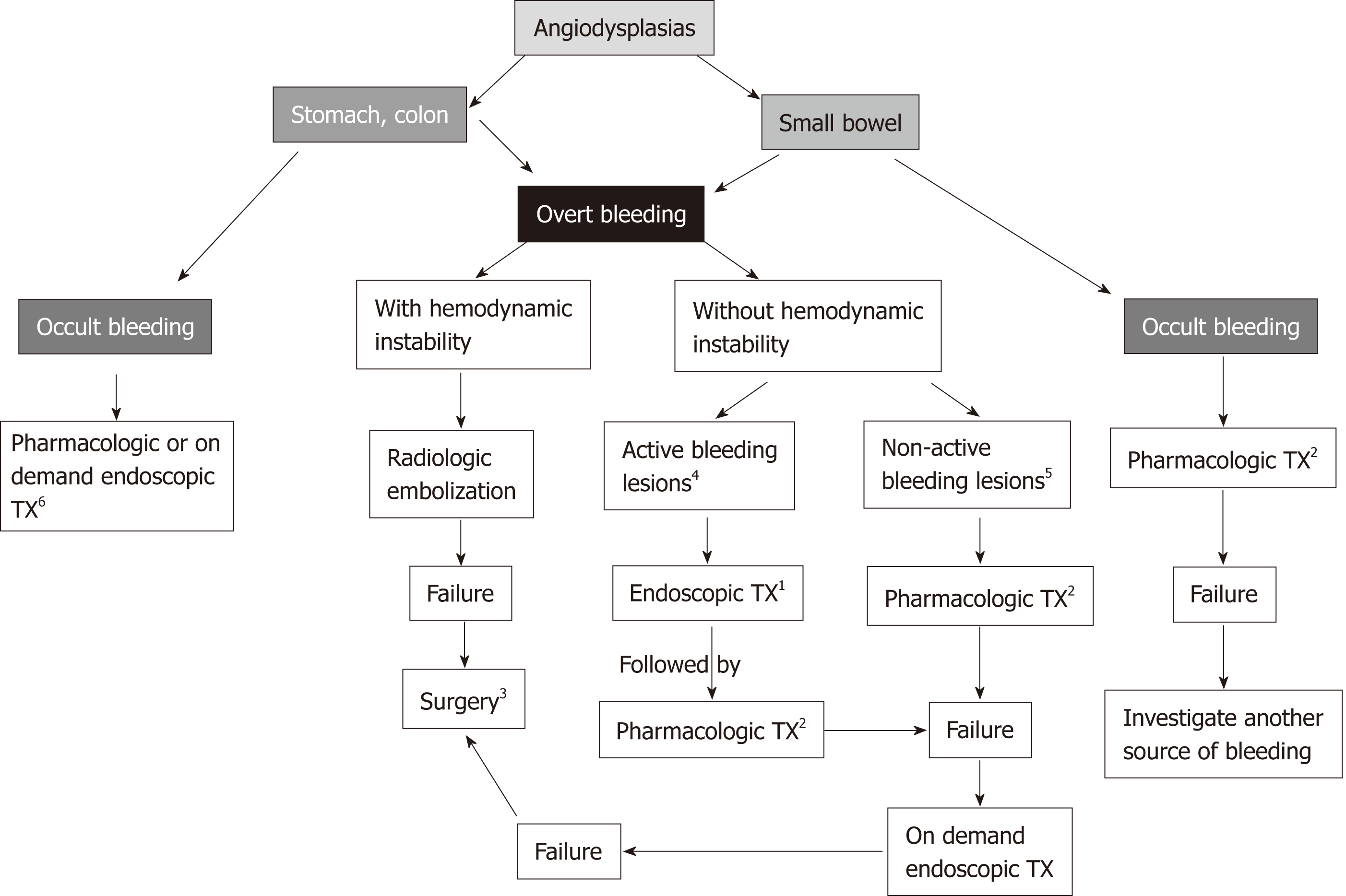
Figure 5 Practical guide for treatment of gastrointestinal angiodysplasias based on their localization, clinical manifestations and endoscopic type lesion.
Small bowel angiodysplasias are separately considered due to their difficult accessibility. 1Hemostatic treatment; 2Prophylactic treatment; 3Consider adequate surgical risk patients with focalized lesions; 4Type 1 (actively bleeding) and Type 2 (with bleeding stigmata); 5Type 3 (bright red spots); 6Alternate use of procedures.
- Citation: García-Compeán D, Del Cueto-Aguilera ÁN, Jiménez-Rodríguez AR, González-González JA, Maldonado-Garza HJ. Diagnostic and therapeutic challenges of gastrointestinal angiodysplasias: A critical review and view points. World J Gastroenterol 2019; 25(21): 2549-2564
- URL: https://www.wjgnet.com/1007-9327/full/v25/i21/2549.htm
- DOI: https://dx.doi.org/10.3748/wjg.v25.i21.2549


