Copyright
©The Author(s) 2019.
World J Gastroenterol. Apr 28, 2019; 25(16): 1936-1949
Published online Apr 28, 2019. doi: 10.3748/wjg.v25.i16.1936
Published online Apr 28, 2019. doi: 10.3748/wjg.v25.i16.1936
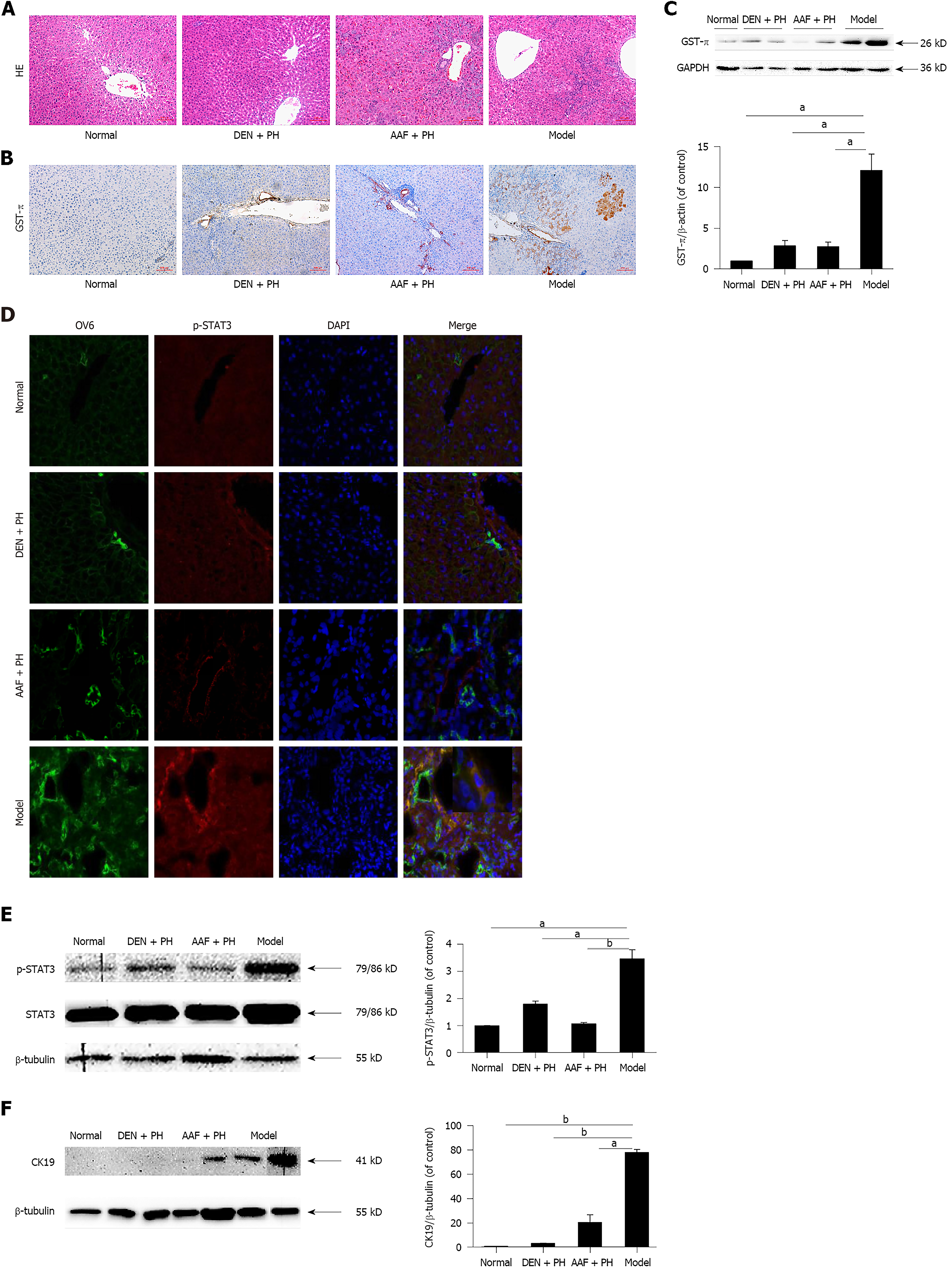
Figure 1 Pathological changes in liver precancerous lesions in rats.
A: Heterocellular clusters were observed in the model group by hematoxylin and eosin staining. Scale bars: 100 μm. B and C: Glutathione S-transferase-π was highly expressed in the model group, as revealed by immunohistochemistry and Western blot. Scale bars: 100 μm. D: Immunofluorescence images showing that p-signal transducer and activator of transcription 3 (STAT3) was highly expressed in activated oval cells in the model group compared with the diethylnitrosamine (DEN) + partial hepatectomy (PH), acetylaminofluorene (AAF) + PH, and normal groups. Scale bars: 50 μm. E: Western blot analysis showed that p-STAT3 was highly expressed in the model group compared with the DEN + PH, AAF + PH, and normal groups. F: Western blot analysis showed that cytokeratin 19 was highly expressed in the model group compared with the DEN + PH, AAF + PH, and normal groups. All values are the mean ± SEM derived from three independent experiments (aP < 0.05, bP < 0.01). GST-π: Glutathione S-transferase-π; STAT3: Signal transducer and activator of transcription 3; CK19: Cytokeratin 19; DEN: Diethylnitrosamine; PH: Partial hepatectomy; AAF: Acetylaminofluorene; HE: Hematoxylin and eosin; OV6: Oval cell 6.
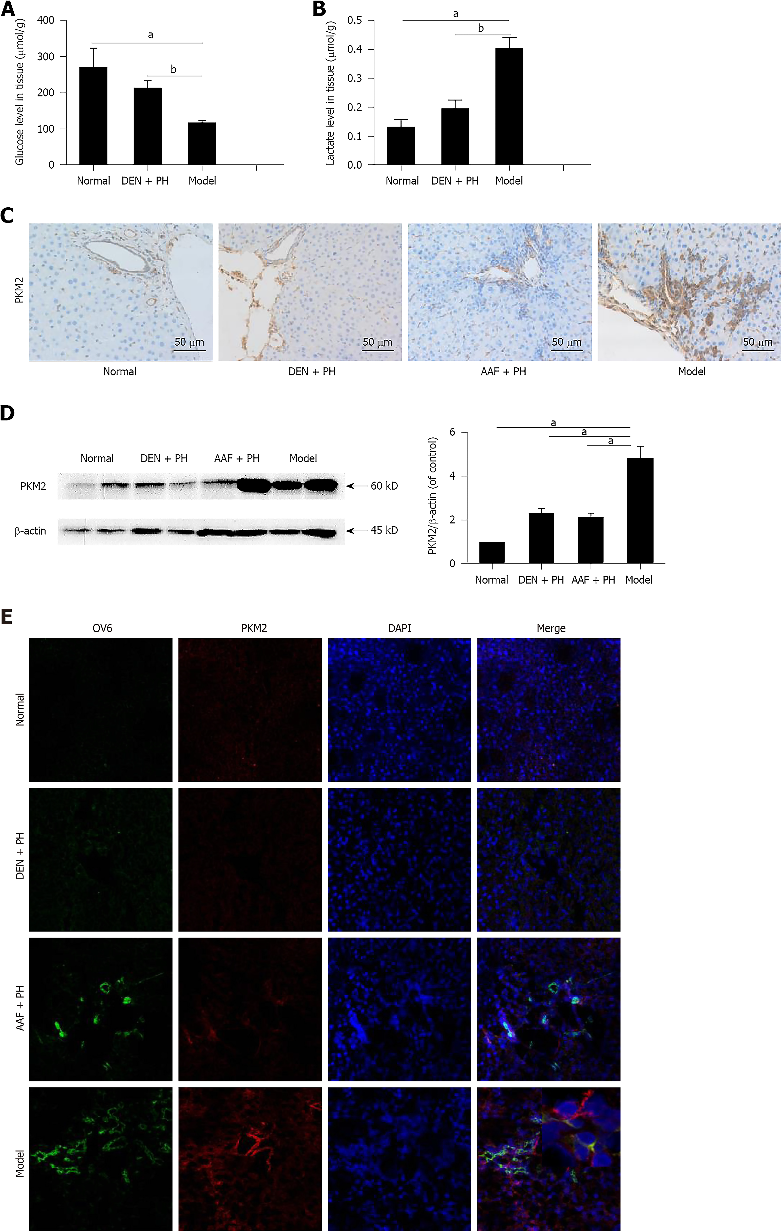
Figure 2 The Warburg effect is initiated in liver precancerous lesions in rats.
A and B: The glucose consumption and lactate production in the model group were elevated compared with those in the diethylnitrosamine (DEN) + partial hepatectomy (PH) and normal groups. C and D: Immunohistochemistry and Western blot analysis showed that pyruvate kinase M2 (PKM2) was highly expressed in the model group compared with that in the DEN + PH, acetylaminofluorene (AAF) + PH, and normal groups. Scale bars: 50 μm. All values are the mean ± SEM derived from three independent experiments (aP < 0.05, bP < 0.01). E: Immunofluorescence images showing that PKM2 was highly expressed in activated oval cells in the model group compared with the DEN + PH, AAF + PH, and normal groups. Scale bars: 75 μm. PKM2: Pyruvate kinase M2; DEN: Diethylnitrosamine; PH: Partial hepatectomy; AAF: Acetylaminofluorene; OV6: Oval cell 6.
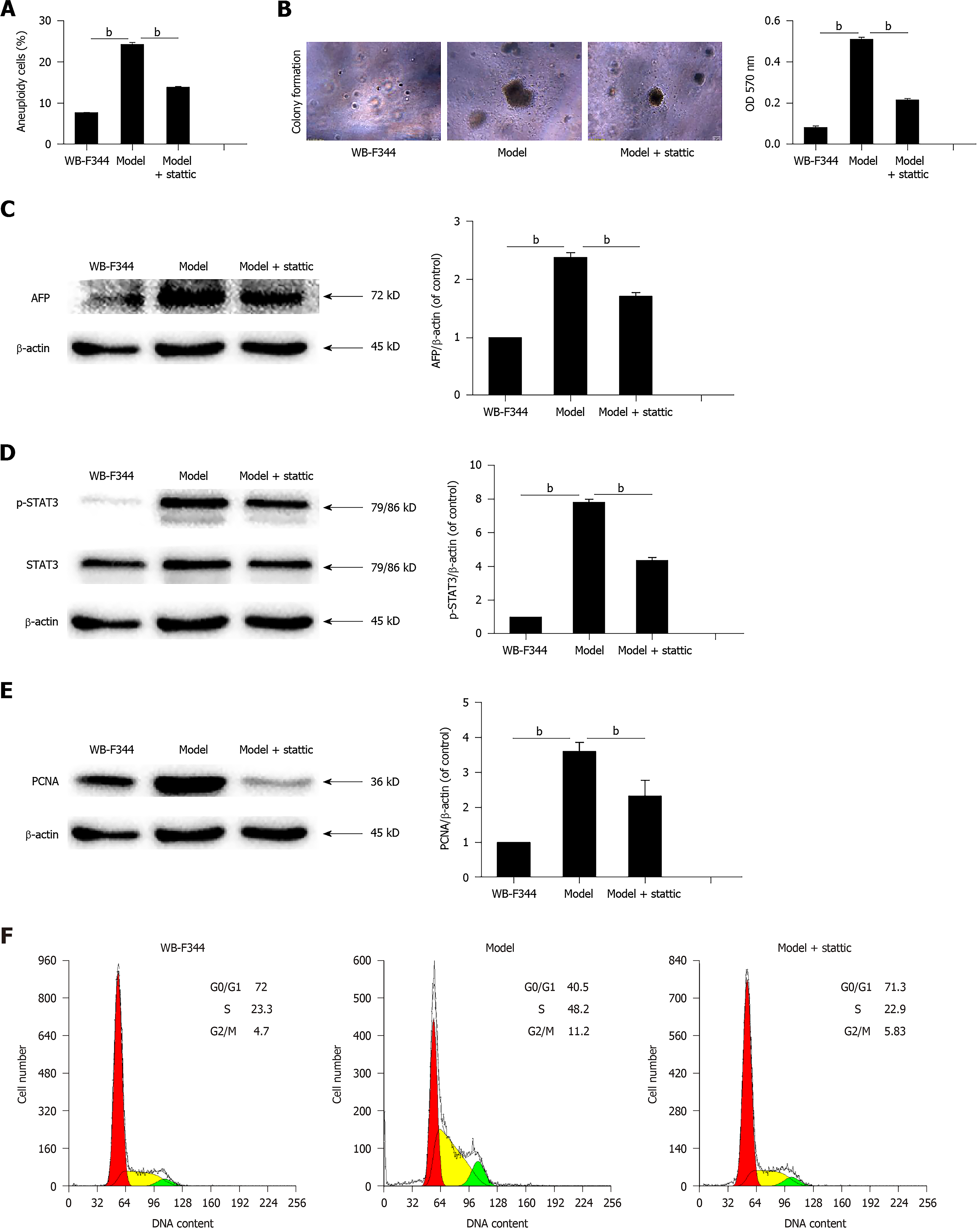
Figure 3 Activated signal transducer and activator of transcription 3 promotes malignant transformation and cell proliferation in WB-F344 cells.
A-D: Aneuploidy, the soft agar colony formation rate, alpha-fetoprotein expression, and p-signal transducer and activator of transcription 3 expression in transformed WB-F344 cells induced with N-methyl-N'-nitro-N-nitroso-guanidine (MNNG) + hydrogen peroxide (H2O2) were increased compared with those in control WB-F344 cells and decreased following stattic treatment. E-G: Proliferating cell nuclear antigen expression and the percentage of cells in S phase in transformed WB-F344 cells induced with MNNG + H2O2 were higher than those in control WB-F344 cells and decreased following stattic treatment. Data are expressed as the mean ± SEM (aP < 0.05, bP < 0.01). (stattic: 4 μmol/L, 48 h). AFP: Alpha-fetoprotein; STAT3: Signal transducer and activator of transcription 3; MNNG: N-methyl-N'-nitro-N-nitroso-guanidine; H2O2: Hydrogen peroxide; PCNA: Proliferating cell nuclear antigen.
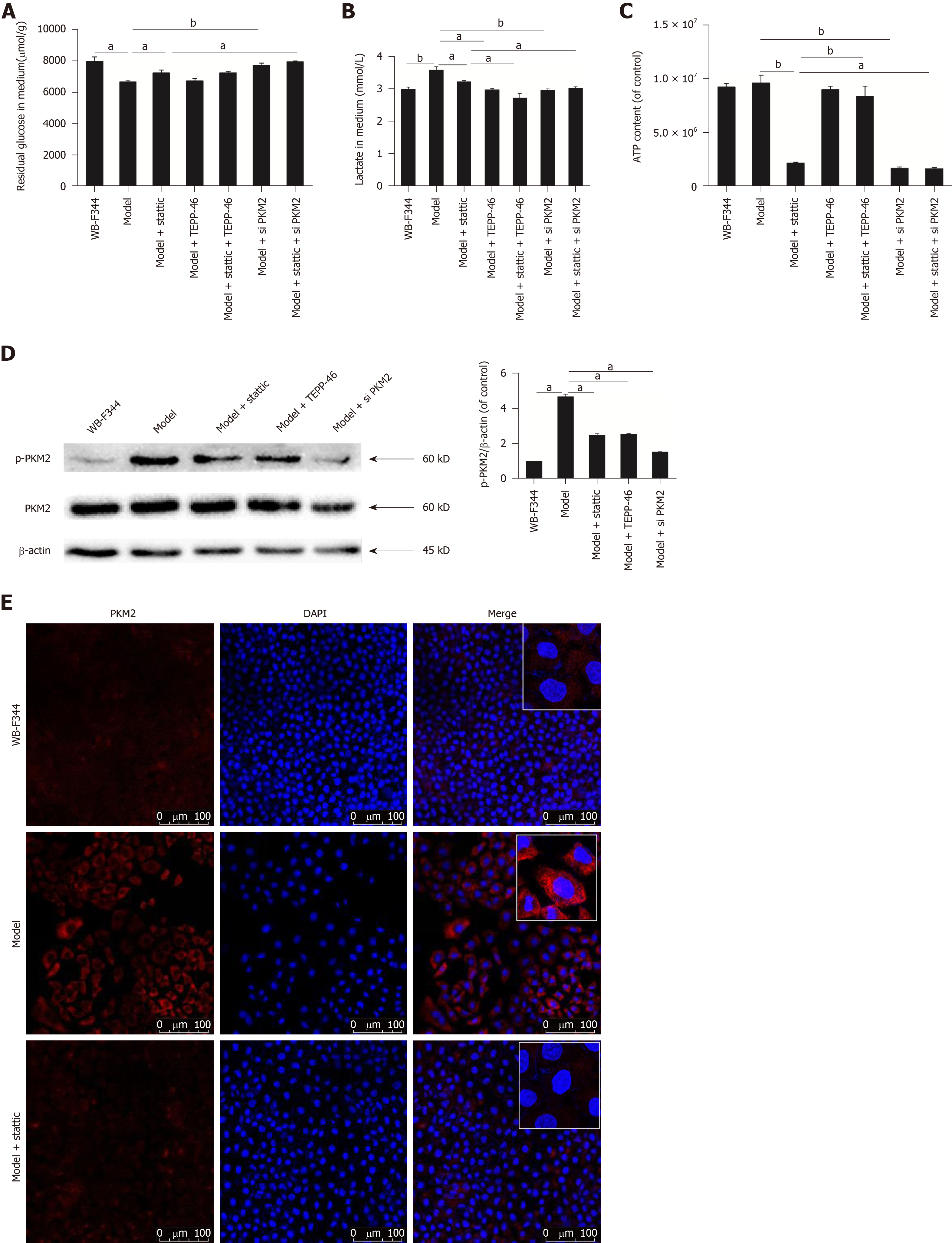
Figure 4 Activated signal transducer and activator of transcription 3 promotes the Warburg effect by enhancing p-pyruvate kinase M2 in transformed WB-F344 cells.
A-C: The glucose concentration in medium in transformed WB-F344 cells induced with N-methyl-N'-nitro-N-nitroso-guanidine (MNNG) + hydrogen peroxide (H2O2) was lower than that in control WB-F344 cells. The lactate concentration in medium in transformed WB-F344 cells induced with MNNG + H2O2 was higher than that in control WB-F344 cells. Inhibition of p-signal transducer and activator of transcription 3 resulted in suppression of glucose consumption, lactate production, and ATP level. TEPP-46 and pyruvate kinase M2 (PKM2) siRNA further reduced the Warburg effect. D: The activation of p-PKM2 was elevated in transformed WB-F344 cells induced with MNNG + H2O2 and was inhibited following stattic treatment. Stattic, TEPP-46, and PKM2 siRNA inhibited the expression of p-PKM2 in transformed WB-F344 cells induced with MNNG + H2O2. E: The expression and nuclear translocation of PKM2 were elevated in transformed WB-F344 cells induced with MNNG + H2O2 and were inhibited following stattic treatment. (aP < 0.05, bP < 0.01) (stattic: 4 μmol/L, 48 h; TEPP-46: 25 μmol/L, 24 h). MNNG: N-methyl-N'-nitro-N-nitroso-guanidine; H2O2: Hydrogen peroxide; STAT3: Signal transducer and activator of transcription 3; PKM2: Pyruvate kinase M2.
- Citation: Bi YH, Han WQ, Li RF, Wang YJ, Du ZS, Wang XJ, Jiang Y. Signal transducer and activator of transcription 3 promotes the Warburg effect possibly by inducing pyruvate kinase M2 phosphorylation in liver precancerous lesions. World J Gastroenterol 2019; 25(16): 1936-1949
- URL: https://www.wjgnet.com/1007-9327/full/v25/i16/1936.htm
- DOI: https://dx.doi.org/10.3748/wjg.v25.i16.1936


