Copyright
©The Author(s) 2019.
World J Gastroenterol. Apr 28, 2019; 25(16): 1913-1927
Published online Apr 28, 2019. doi: 10.3748/wjg.v25.i16.1913
Published online Apr 28, 2019. doi: 10.3748/wjg.v25.i16.1913
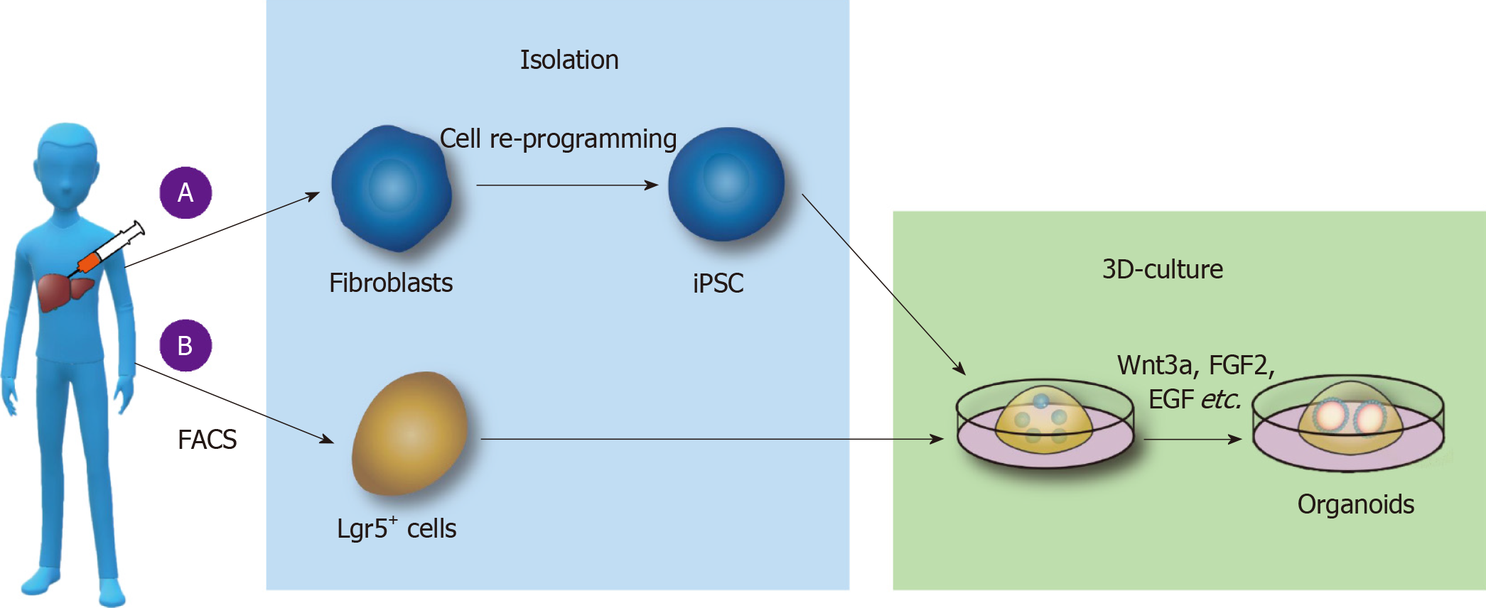
Figure 1 Culture of liver organoids in vitro.
A: Liver organoids cultured from pluripotent stem cells (PSCs). Somatic cells are reprogrammed into induced PSCs; epidermal growth factor (EGF), fibroblast growth factor 2 (FGF2), and other substances are added to the medium; and the cells are cultured on Matrigel to form liver organoids; B: Liver organoids cultured from adult stem cells. Lgr5+ cells are isolated by fluorescence-activated cell sorting, and Wnt3a, Y27632, EGF, Rspo1, FGF, and other substances are added to form organoids by 3D culture. FACS: Fluorescence-activated cell sorting; iPSCs: Induced pluripotent stem cells; FGF2: Fibroblast growth factor 2; EGF: Epidermal growth factor.
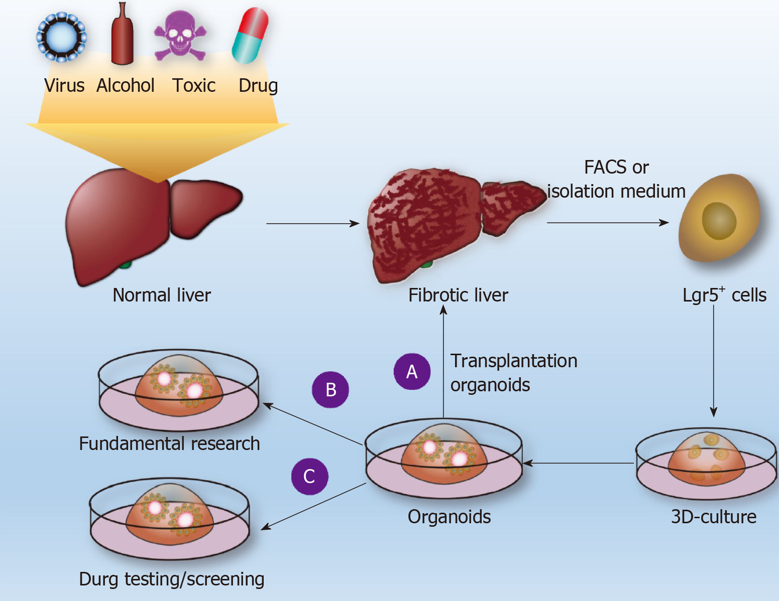
Figure 2 Use of liver organoids to study liver fibrosis.
Normal liver tissue is subjected to pathogenic factors (viruses, alcohol, drugs, poisons, etc.) to form liver fibrosis (LF), and Lgr5+ cells are obtained by fluorescence-activated cell sorting or conventional separation to form individualized liver organoids. A: Such organoids are used for transplantation to restore liver function; B: Basic research on LF; C: Drug sensitivity tests. FACS: Fluorescence-activated cell sorting.
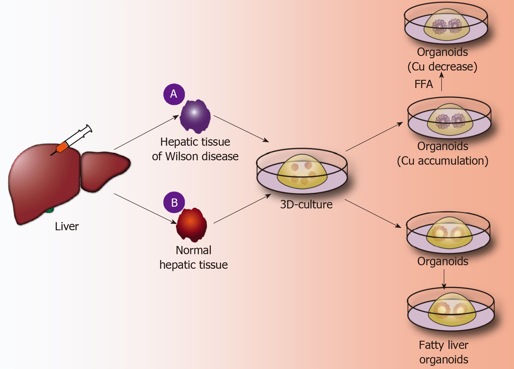
Figure 3 Application of liver organoids in benign hepatic diseases.
A: A fine needle is used to puncture Wilson disease tissue and other genetically deficient liver tissues; 3D organoid culture technology is used to obtain the corresponding gene-deficient liver organoids, copper accumulation is identified inside the organoids, and the further using a CRISPR/Cas9 system is used to correct defective genes, thereby significantly reducing copper accumulation in liver organoids; B: A fine needle is used to puncture normal liver tissue, and normal liver organoids are obtained by the 3D organoid culture technique. Free fatty acids are added to the culture medium, and the liver cells in the organoids show lipid accumulation. FFA: Free fatty acids.
Figure 4 Culture and application of liver cancer organoids.
A: Culture of liver cancer organoids. Primary liver cancer cells are obtained after the enzymatic hydrolysis of collagenase, and similar liver organoids are used to form liver cancer organoids; B: Malignant transformation of normal liver organoids to form liver cancer organoids. Liver organoids are routinely formed, gene editing technology is used to generate multiple-gene, multistep mutations in organoids, and the organoids are orderly cultured to form liver cancer organoids; C: Application of liver cancer organoids. Liver cancer organoids can be used for research of basic mechanism of liver cancer and screening of liver cancer drugs, for example, screening the best chemotherapeutic drugs by comparing the effects of different drugs on the size or number of liver cancer organoids. HCC: Hepatocellular carcinoma; FGF2: Fibroblast growth factor 2; EGF: Epidermal growth factor.
- Citation: Wu LJ, Chen ZY, Wang Y, Zhao JG, Xie XZ, Chen G. Organoids of liver diseases: From bench to bedside. World J Gastroenterol 2019; 25(16): 1913-1927
- URL: https://www.wjgnet.com/1007-9327/full/v25/i16/1913.htm
- DOI: https://dx.doi.org/10.3748/wjg.v25.i16.1913


