Copyright
©The Author(s) 2019.
World J Gastroenterol. Apr 14, 2019; 25(14): 1775-1782
Published online Apr 14, 2019. doi: 10.3748/wjg.v25.i14.1775
Published online Apr 14, 2019. doi: 10.3748/wjg.v25.i14.1775
Figure 1 Clinical picture and imaging materials of the patient.
A: The enteroatmospheric fistula (EAF) happened at side-to-side anastomosis was exposed and visible after open abdomen (red arrow); B: Contrast-mediated fistula angiography through the orifice of the fistulous tract (red arrow); C: The location of initial obstruction was shown (red arrow).
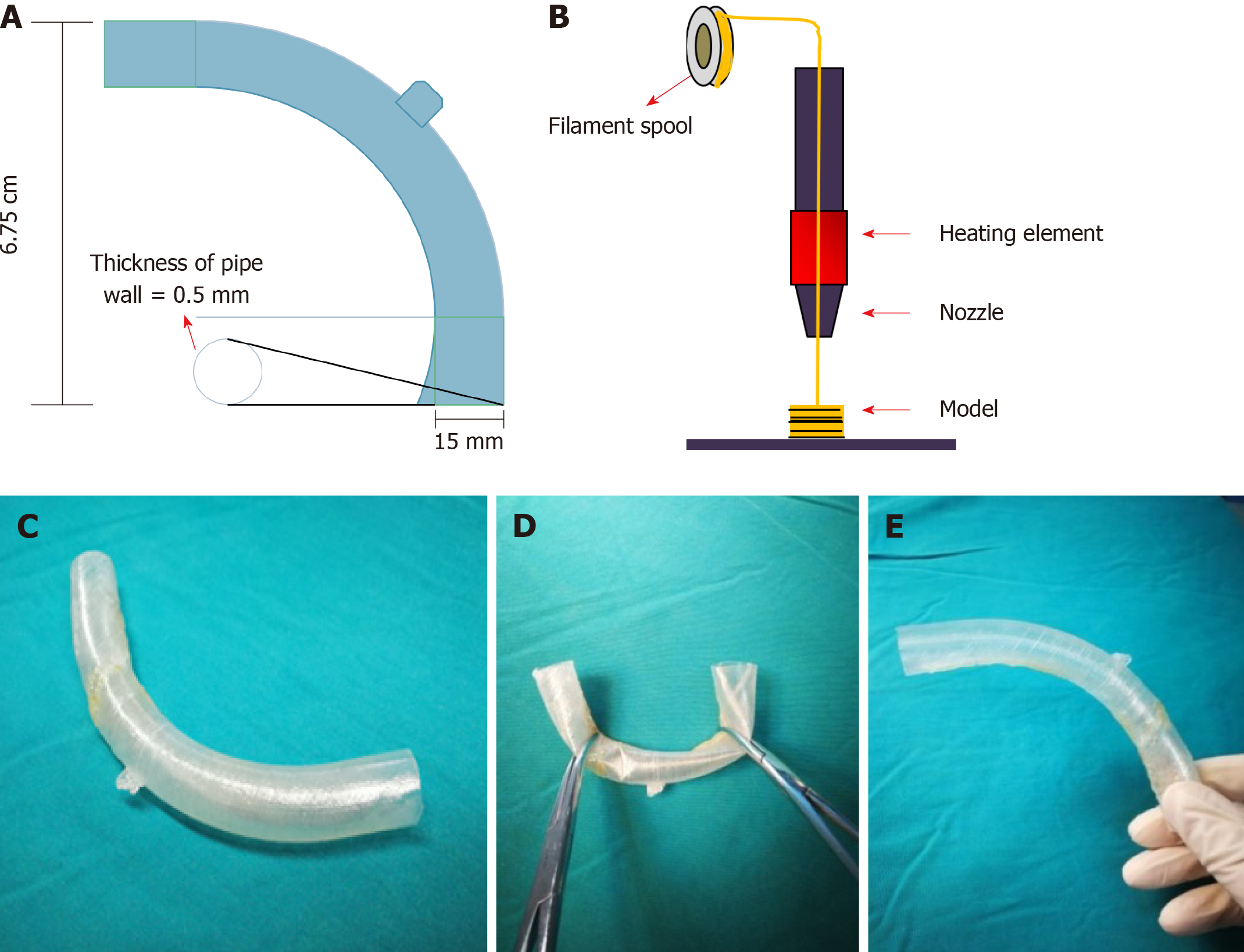
Figure 2 Manufacturing process of the “fistula stent”.
A: The blueprint of “fistula stent” was built with Solidwork software and saved in Standard Template Library (STL) form; B: Fused Deposition Modeling is a process where thermoplastic filaments are driven with a motorized heated nozzle; C: Build model of a hollow and curving pipe stent with a small protuberance and integrated wall; D: Flexibility of the “fistula stent”; E: Shape memory of the “fistula stent” after application of an external force.
Figure 3 Process of the implantation and fixation of the “fistula stent”.
A: Four double pipes were used to drainage enteric fistula effluent before implantation ; B: The process of implantation; C: After the implantation the stent can still be seen (red arrow); D: The process of fixation by suturing the small protuberance with a latex tube overhanging the abdominal wall; E: After the accomplishment of implantation, only one double pipe was needed for drainage; F: Contrast-mediated fistula angiography was conducted to verify the effectiveness of the stent and the contrast agent flowed past the stent without obstruction.
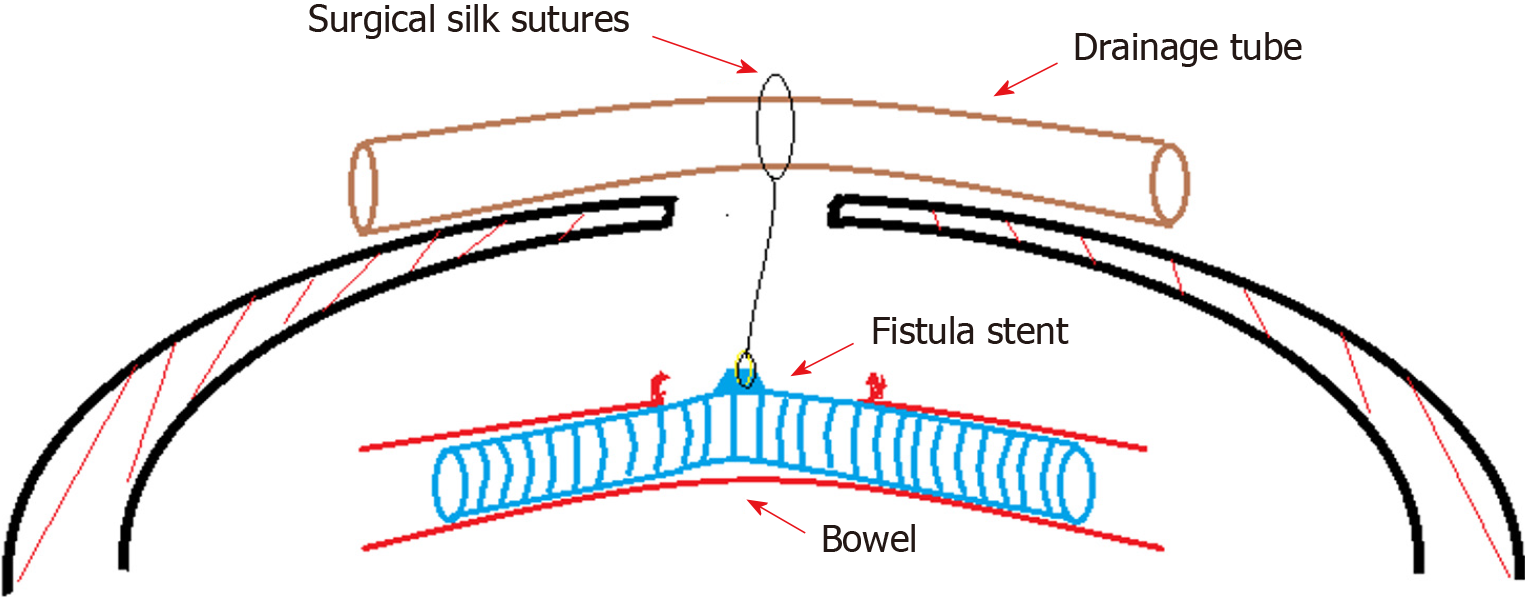
Figure 4 Concept graph of how is the “fistula stent” fixed in the bowel.
The protuberance is connected with a latex drainage tube hanging over the abdominal wall in order to prevent the sliding due to peristalsis.
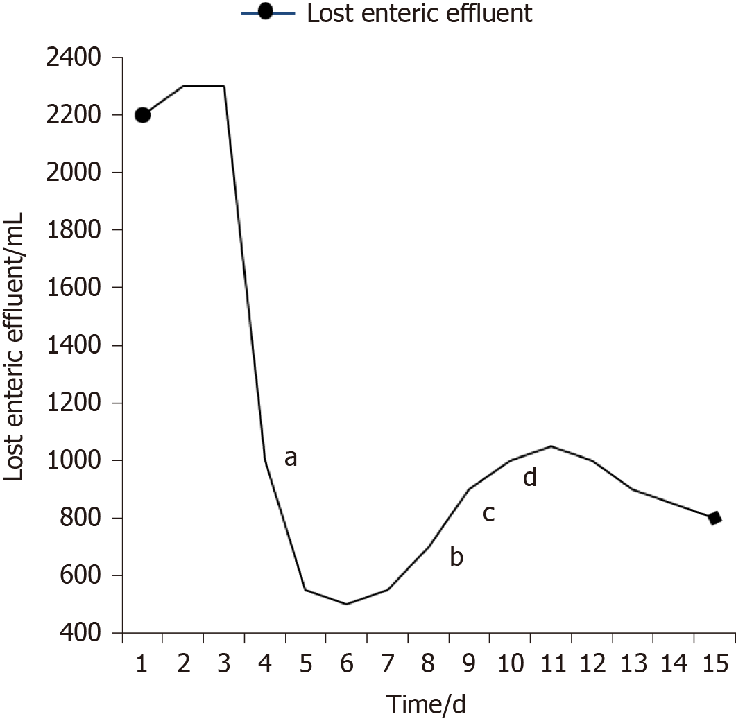
Figure 5 Recorded amount of lost enteric effluent.
aThe day of implantation; bStarting enteral nutrition (EN) of 500 mL; cEN of 1000 mL; dEN of 1500 mL.
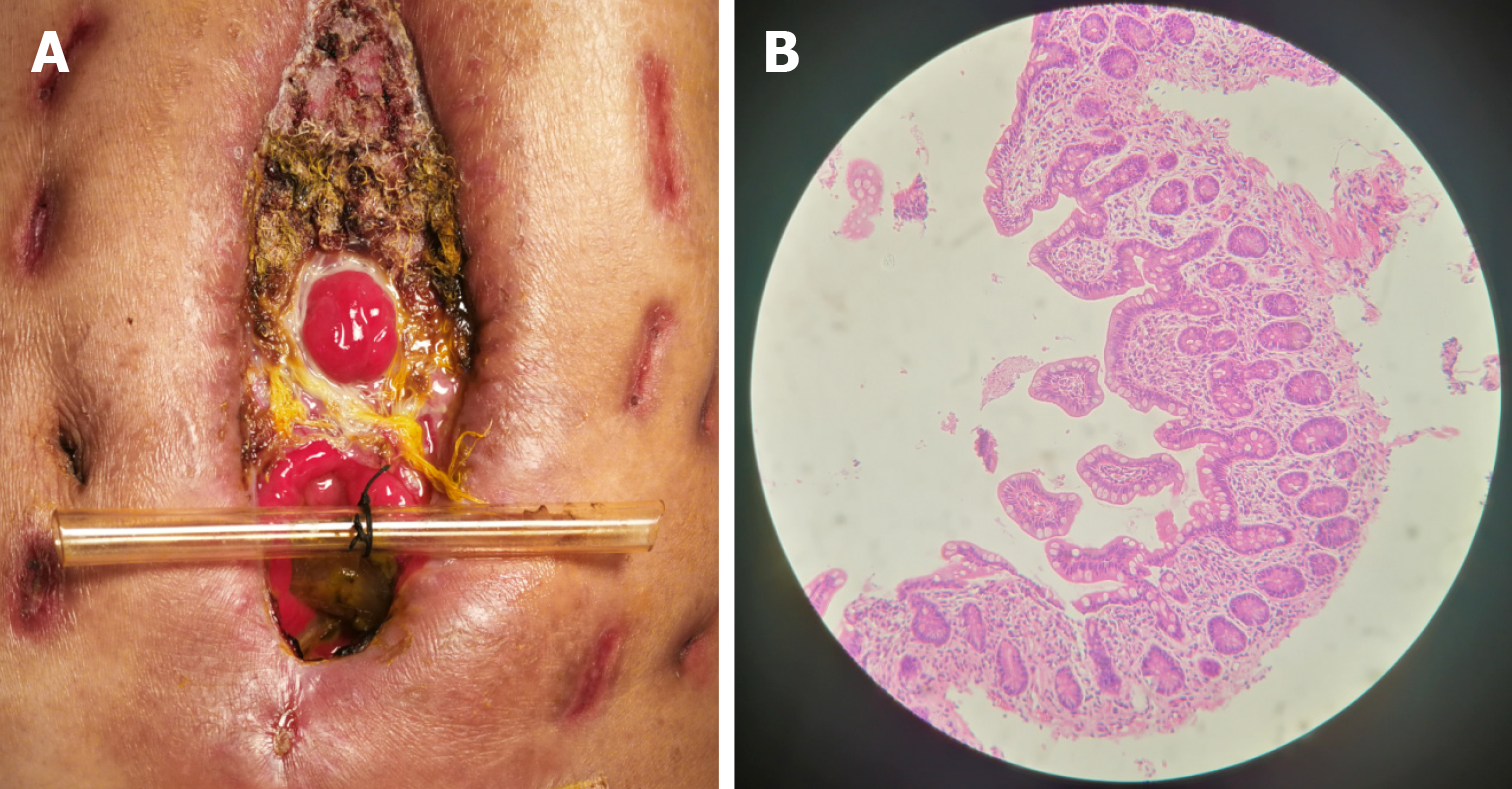
Figure 6 Follow-up after the implantation.
A: After skin grafting, the abdomen had granulated around the enteroatmospheric fistula (EAF) and the orifice of the fistulous tract had also decreased to a very small size; B: Atrophic intestinal mucosa in the distal end of the EAF before restoring enteral nutrition.
- Citation: Xu ZY, Ren HJ, Huang JJ, Li ZA, Ren JA. Application of a 3D-printed ”fistula stent” in plugging enteroatmospheric fistula with open abdomen: A case report. World J Gastroenterol 2019; 25(14): 1775-1782
- URL: https://www.wjgnet.com/1007-9327/full/v25/i14/1775.htm
- DOI: https://dx.doi.org/10.3748/wjg.v25.i14.1775


