Copyright
©The Author(s) 2019.
World J Gastroenterol. Apr 14, 2019; 25(14): 1684-1696
Published online Apr 14, 2019. doi: 10.3748/wjg.v25.i14.1684
Published online Apr 14, 2019. doi: 10.3748/wjg.v25.i14.1684
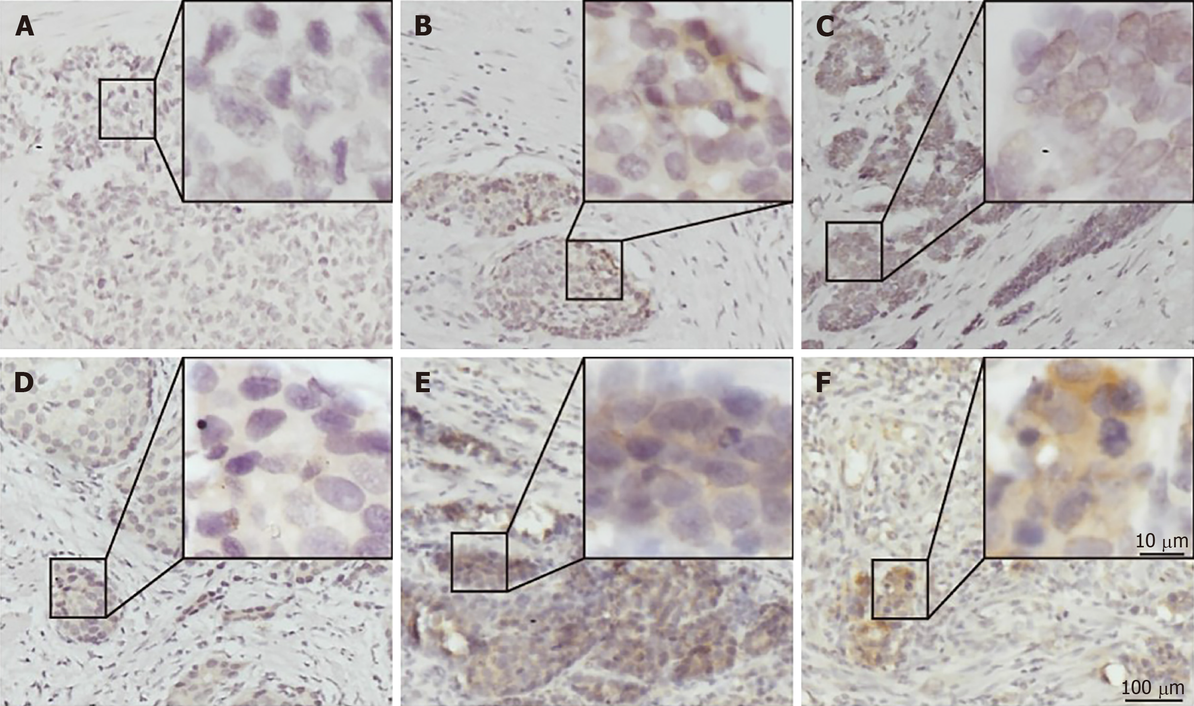
Figure 1 Immunohistochemical staining for programmed death ligand 1 in human gastric neuroendocrine carcinomas.
A: Representative case of negative programmed death ligand 1 (PD-L1) expression; B: Tumor-stromal interface enhanced expression of PD-L1; C: Membrane expression of PD-L1; D: Weak staining; E: Moderate staining; F: Strong staining. PD-L1: Programmed death ligand 1.
Figure 2 Immunohistochemical staining for programmed death 1, CD8, and FOXP3 in human gastric neuroendocrine carcinomas.
A: The programmed death 1 positive infiltrating lymphocytes; B: CD8 positive lymphocytes; C: FOXP3 positive Treg cells.
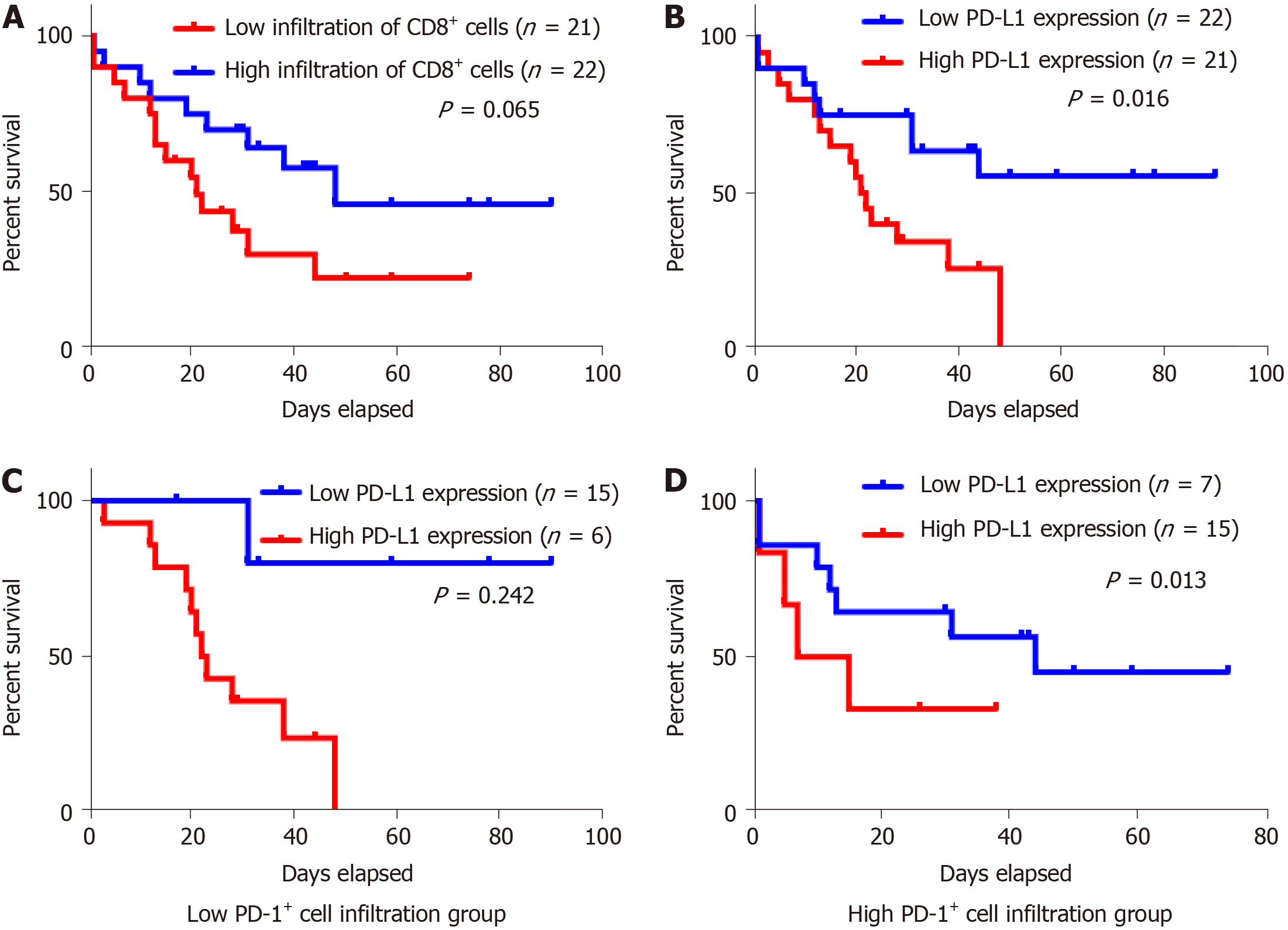
Figure 3 Kaplan–Meier survival analysis of the gastric neuroendocrine carcinomas patients.
A: Patients with high infiltration of CD8+ lymphocytes tended to have a relative longer survival, but the difference was not statistically significant; B: Kaplan-Meier overall survival curves in patients with high and low expression of programmed death ligand 1 (PD-L1) in the whole population; C: Low PD-1+ cell infiltration group; D: High PD-1+ cell infiltration group. PD-L1: Programmed death ligand 1.
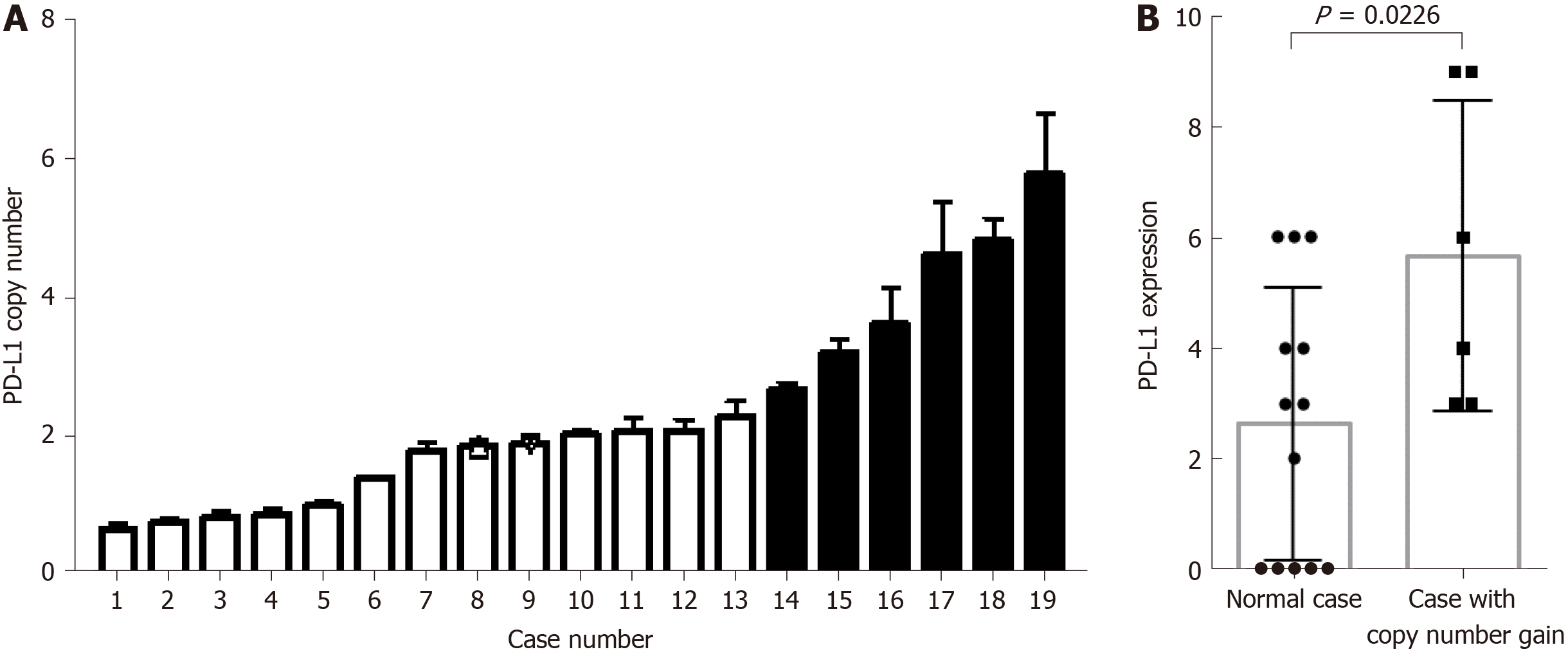
Figure 4 Programmed death ligand 1 copy number is associated with programmed death ligand 1 expression in gastric neuroendocrine carcinomas.
A: Programmed death ligand 1 (PD-L1) copy number status varied in 19 gastric neuroendocrine carcinoma samples. The cases with copy number gain are shown in black; B: Cases with copy number gain showed significantly higher PD-L1 expression than those without. PD-L1: Programmed death ligand 1.
- Citation: Yang MW, Fu XL, Jiang YS, Chen XJ, Tao LY, Yang JY, Huo YM, Liu W, Zhang JF, Liu PF, Liu Q, Hua R, Zhang ZG, Sun YW, Liu DJ. Clinical significance of programmed death 1/programmed death ligand 1 pathway in gastric neuroendocrine carcinomas. World J Gastroenterol 2019; 25(14): 1684-1696
- URL: https://www.wjgnet.com/1007-9327/full/v25/i14/1684.htm
- DOI: https://dx.doi.org/10.3748/wjg.v25.i14.1684


