Copyright
©The Author(s) 2019.
World J Gastroenterol. Apr 14, 2019; 25(14): 1653-1665
Published online Apr 14, 2019. doi: 10.3748/wjg.v25.i14.1653
Published online Apr 14, 2019. doi: 10.3748/wjg.v25.i14.1653
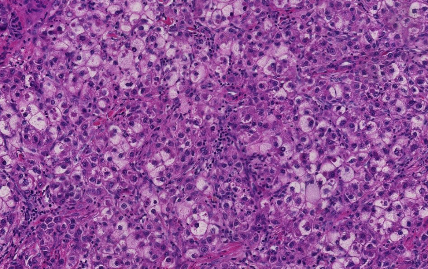
Figure 1 Steatohepatitic hepatocellular carcinoma.
Tumoral cells show features of steatohepatitis including steatosis, ballooning and Mallory-Denk bodies (Hematoxylin and eosin stain, × 200).
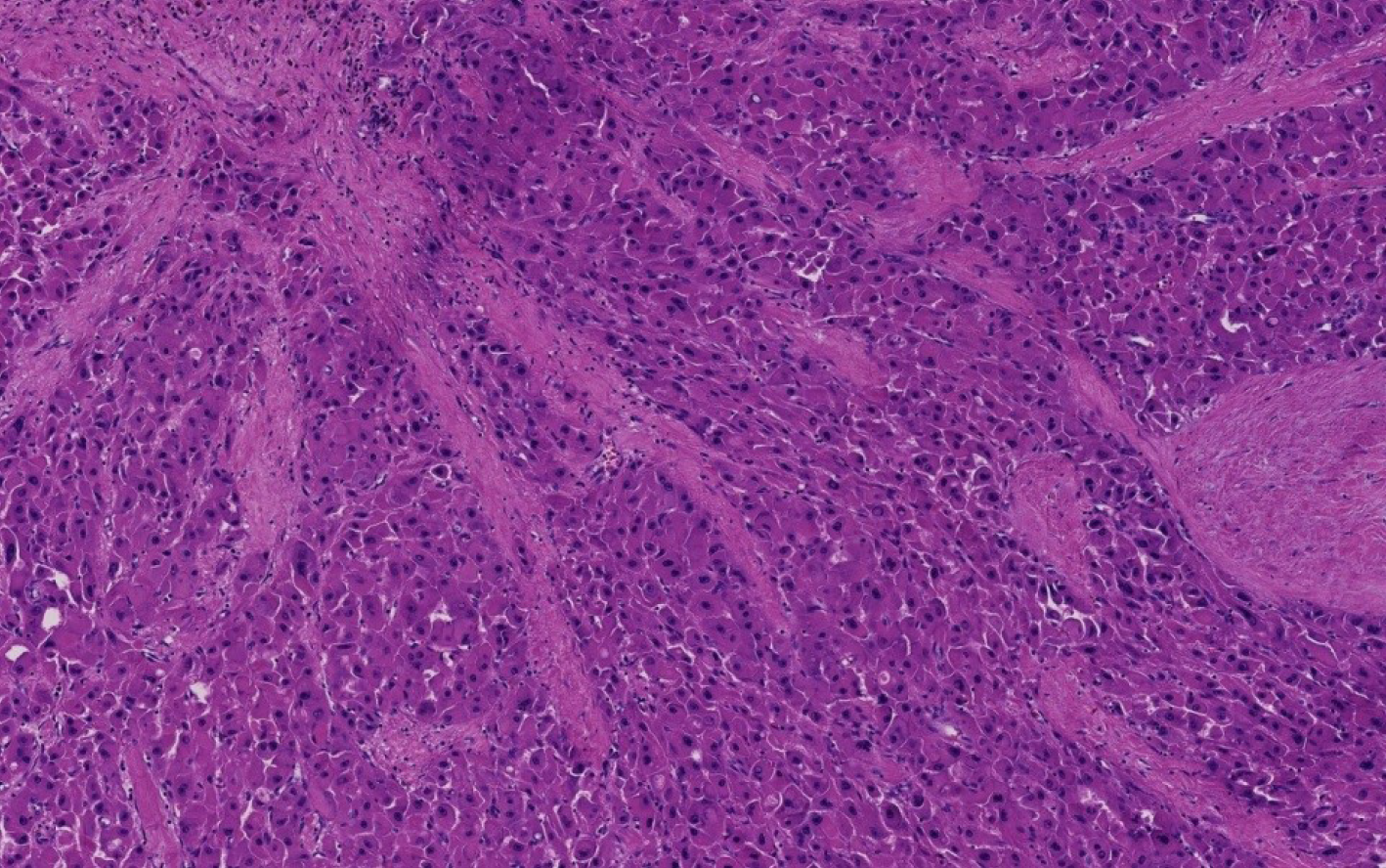
Figure 2 Fibrolamellar hepatocellular carcinoma.
Multiple fibrous septa composed of parallel collagen fibers separate tumoral trabeculae (Hematoxylin and eosin stain, × 100).
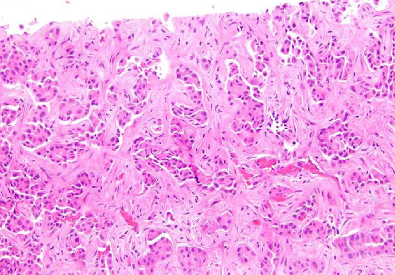
Figure 3 Scirrhous hepatocellular carcinoma.
Bands of dense fibrosis separate cords and nests of tumoral cells (Hematoxyin and eosin stain, × 200).
Figure 4 Combined hepatocellular carcinoma-cholangiocarcinoma.
Areas of conventional hepatocellular carcinoma (lower mid to right) are admixed with areas of discrete glands formation (upper left) (Hematoxylin and eosin stain, × 40).
- Citation: El Jabbour T, Lagana SM, Lee H. Update on hepatocellular carcinoma: Pathologists’ review. World J Gastroenterol 2019; 25(14): 1653-1665
- URL: https://www.wjgnet.com/1007-9327/full/v25/i14/1653.htm
- DOI: https://dx.doi.org/10.3748/wjg.v25.i14.1653


