Copyright
©The Author(s) 2019.
World J Gastroenterol. Jan 7, 2019; 25(1): 85-94
Published online Jan 7, 2019. doi: 10.3748/wjg.v25.i1.85
Published online Jan 7, 2019. doi: 10.3748/wjg.v25.i1.85
Figure 1 Surgery of celiac trunk ganglion damage.
A: The aorta ventralis was observed in the endoscopic direct status and the aortic tissue was clamped by thermal biopsy forceps; B: The peritoneal tissue was partially removed by biopsy forceps for simulating ganglion destruction; C: Anatomic structure in the posterior gastric wall. The white arrow shows the lesser omental sac, and the two yellow loops show tissue adhesion.
Figure 2 Surgery of partial hepatectomy.
A and B: Partial hepatectomy using an electric knife; C: The wound in the left hepatic lobe; D: Hepatic tissue stained with hematoxylin and eosin (200 ×).
Figure 3 Surgery of partial splenectomy.
A: Partial splenectomy using electric knife; B: Wedge resection of the spleen and massive bleeding during operation; C: The wound in the spleen tail; D: Spleen tissue stained with hematoxylin and eosin (200 ×).
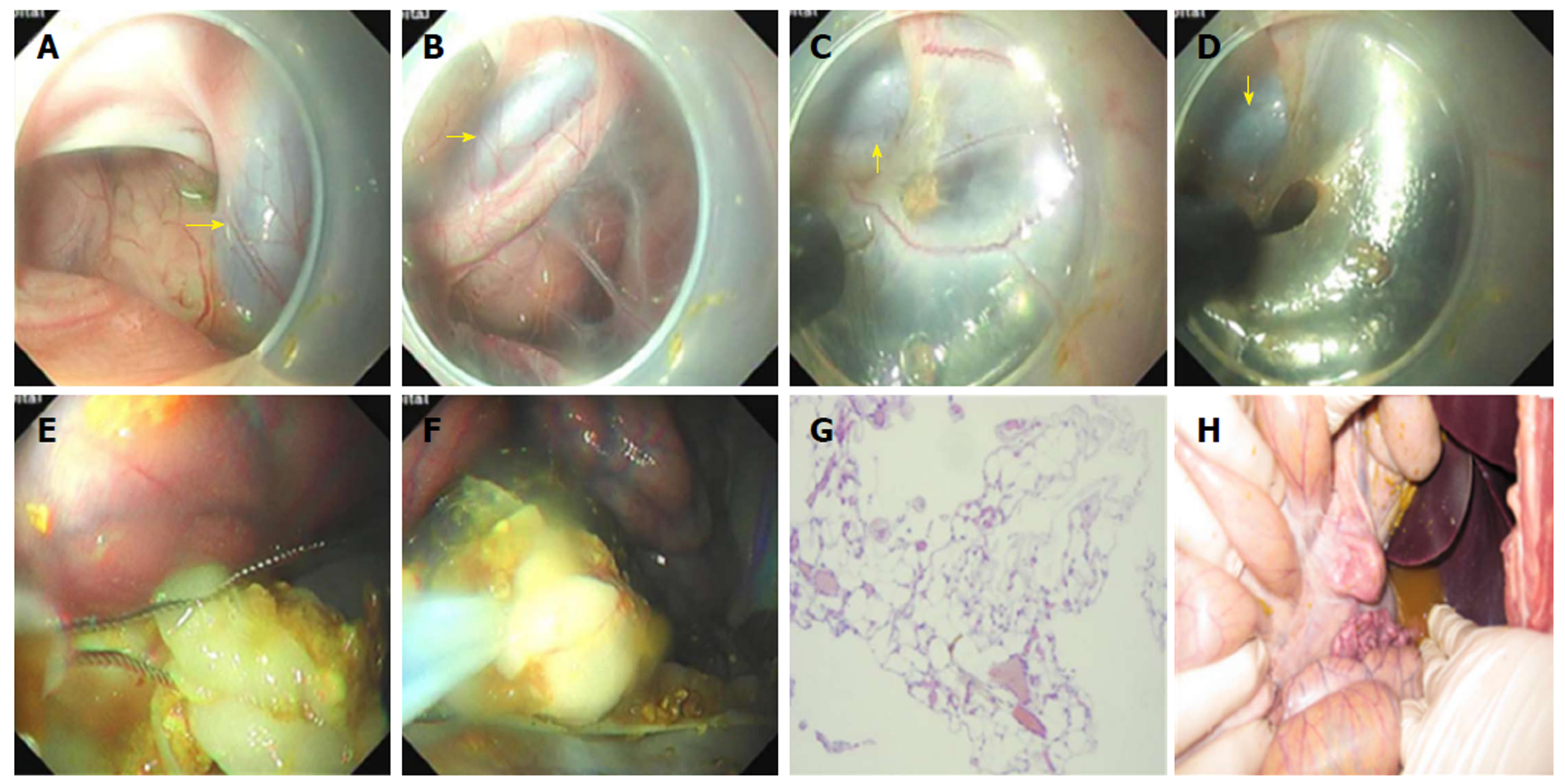
Figure 4 Surgery of retroperitoneal tissue resection.
A: The aorta ventralis was observed in the endoscopic direct status; B: The retroperitoneal space was visible near the aorta ventralis; C and D: Partial resection of tissues in the retroperitoneal space using an electric knife; E and F: Partial resection of the omentum majus using an endoloop; G: Omentum majus tissue stained with hematoxylin and eosin (200 ×); H: A large amount of liquid and chyme in the abdominal cavity.
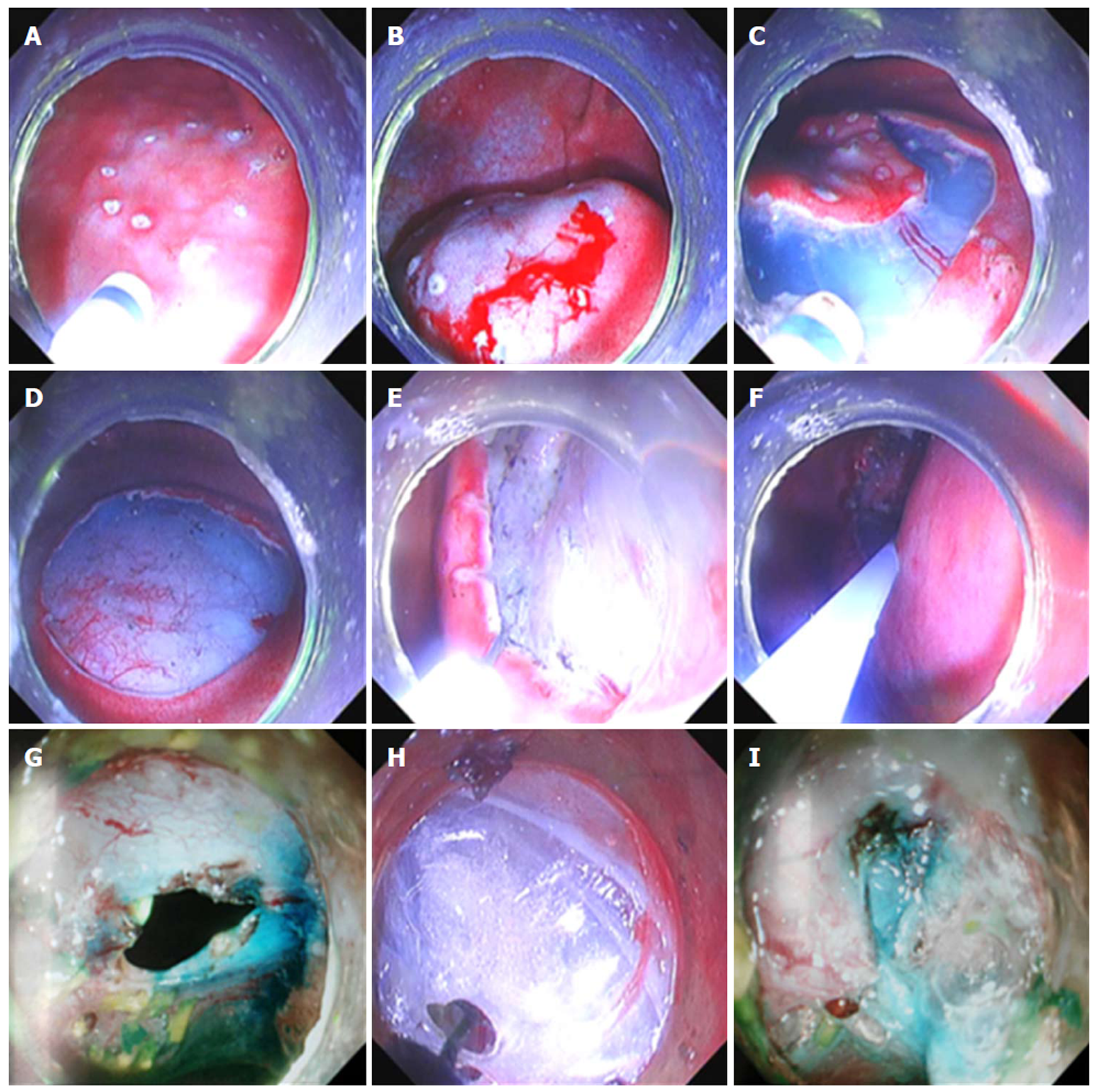
Figure 5 Surgery of endoscopic submucosal dissection and lymph node dissection.
A-D: Simulation of ESD of early gastric carcinoma in pigs; E and F: Methylene blue marker injected to the intrinsic muscle layer surrounding the wound; G and H: Dissection of lymph nodes outside the gastric wall; G: Incision of the serosa and muscularis propria; H: Separation of tissues outside the gastric wall; I: A close large gastric perforation using two endoscopes. ESD: Endoscopic submucosal dissection.
- Citation: Xiong Y, Chen QQ, Chai NL, Jiao SC, Ling Hu EQ. Endoscopic trans-esophageal submucosal tunneling surgery: A new therapeutic approach for diseases located around the aorta ventralis. World J Gastroenterol 2019; 25(1): 85-94
- URL: https://www.wjgnet.com/1007-9327/full/v25/i1/85.htm
- DOI: https://dx.doi.org/10.3748/wjg.v25.i1.85


