Copyright
©The Author(s) 2018.
World J Gastroenterol. Jan 28, 2018; 24(4): 475-483
Published online Jan 28, 2018. doi: 10.3748/wjg.v24.i4.475
Published online Jan 28, 2018. doi: 10.3748/wjg.v24.i4.475
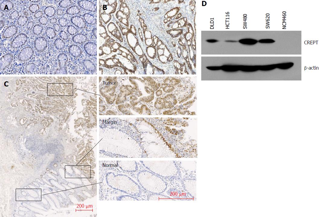
Figure 1 CREPT expression in tumor and adenoma tissues.
A: Negative CREPT immunohistochemical staining in colorectal adenoma tissue; B: Positive CREPT immunohistochemical staining in CRC tissue; C: CREPT expression pattern in CRC sample; D: CREPT was determined by immunoblotting in CRC cell lines. CRC: Colorectal carcinoma.
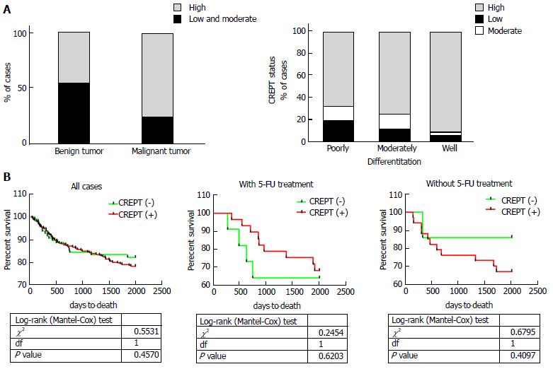
Figure 2 CREPT expression in CRC correlates with clinicopathological characteristics.
A: CREPT expression level correlates with pathological type and tumor differentiation; B: Survival curve of CRC patients shows significant difference between patients with or without 5-FU-bases adjuvant chemotherapy. CRC: Colorectal carcinoma.
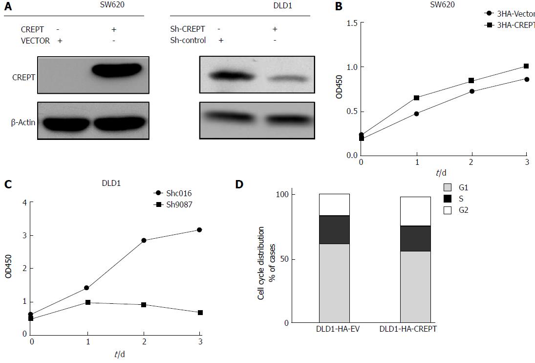
Figure 3 Alteration of CREPT expression in CRC cells affects cell proliferation.
A: Western blotting showed that expression of CREPT in SW620 and DLD1 changed after exposed to virus for 48 h; B and C: SW620 and DLD1 cells were incubated in 96-well plates, and CCK-8 assay determined cell viability; D: Cell cycle was detected by flow cytometry and indicated that CREPT accelerated cells through the G1/S check point. CRC: Colorectal carcinoma.
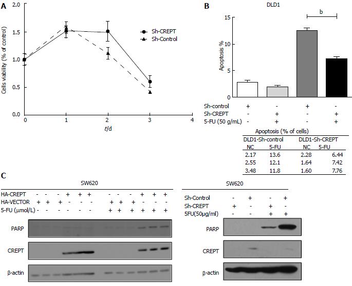
Figure 4 CREPT facilitates CRC cell response to 5-FU chemotherapy.
A: Cells were treated with 5-FU (50 μg/mL), and CCK-8 assay estimated their viability; B: Annexin V-PI apoptosis detection show that DLD1 cells were less sensitive to 5-FU chemotherapy when CREPT was knocked down; C: SW620 and DLD1 cells were harvested after exposure to 5-FU (50 μg/mL) for 48 h, and western blotting showed that PARP was highly expressed in cells that expressed more CREPT. CRC: Colorectal carcinoma.
- Citation: Kuang YS, Wang Y, Ding LD, Yang L, Wang Y, Liu SH, Zhu BT, Wang XN, Liu HY, Li J, Chang ZJ, Wang YY, Jia BQ. Overexpression of CREPT confers colorectal cancer sensitivity to fluorouracil. World J Gastroenterol 2018; 24(4): 475-483
- URL: https://www.wjgnet.com/1007-9327/full/v24/i4/475.htm
- DOI: https://dx.doi.org/10.3748/wjg.v24.i4.475


