Copyright
©The Author(s) 2018.
World J Gastroenterol. Sep 21, 2018; 24(35): 4061-4068
Published online Sep 21, 2018. doi: 10.3748/wjg.v24.i35.4061
Published online Sep 21, 2018. doi: 10.3748/wjg.v24.i35.4061
Figure 1 Representative endoscopic images of patients in each of the groups.
A and B: Group A (serum antibody titer < 3 U/mL). A 20-year-old woman with a serum antibody titer < 3 U/mL and Kyoto classification score of 0. Atrophy was absent and RAC and superficial gastritis were present; C and D: Group C (serum antibody titer of 10-49.9 U/mL). A 36-year-old woman with a serum antibody titer of 25.5 U/mL and Kyoto classification score of 1. Closed-type atrophy was present and enlarged folds, nodularity, diffuse redness, and RAC were absent; E and F: Group D (serum antibody titer ≥ 50 U/mL). A 50-year-old woman with a serum antibody titer ≥ 100 U/mL and Kyoto classification score of 7. Open-type atrophy, enlarged folds, nodularity, and diffuse redness were present, and RAC was absent. RAC: Regular arrangement of collecting venules.
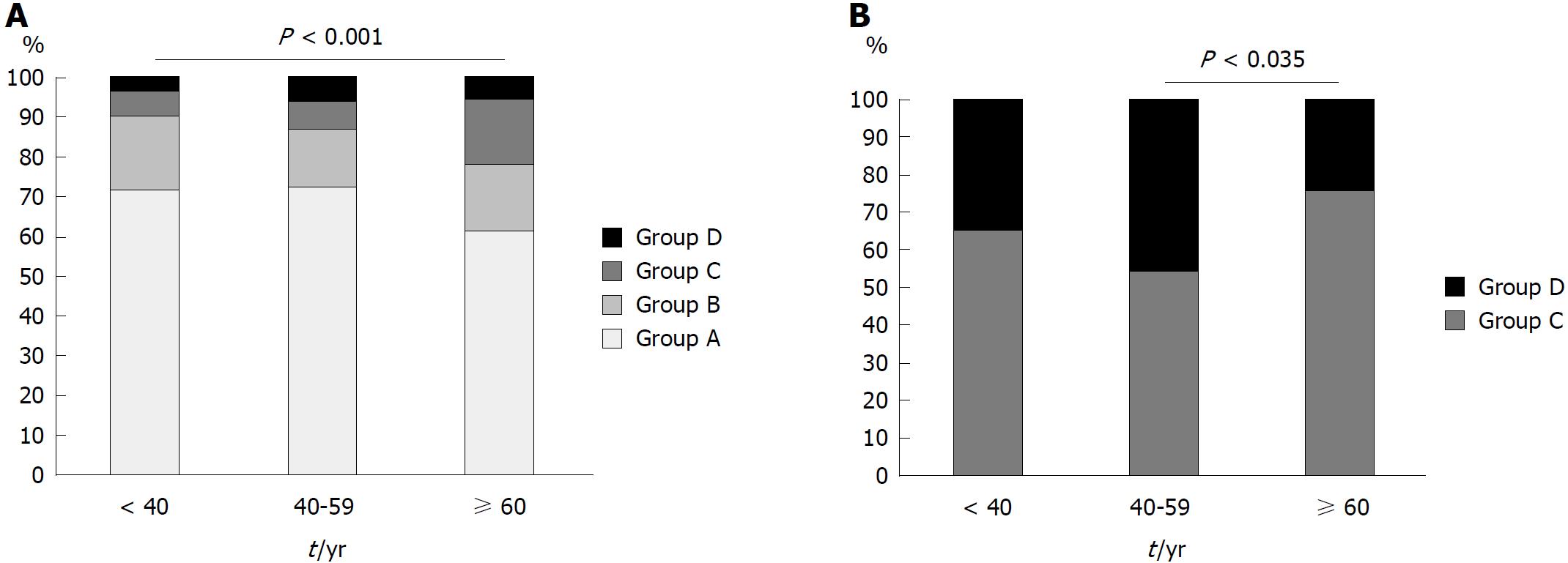
Figure 2 Proportion of Helicobacter pylori-infected patients.
A: The proportion of patients in each of the groups, stratified by age. B: The proportion of patients in groups C and D, stratified by age.
- Citation: Toyoshima O, Nishizawa T, Sakitani K, Yamakawa T, Takahashi Y, Yamamichi N, Hata K, Seto Y, Koike K, Watanabe H, Suzuki H. Serum anti-Helicobacter pylori antibody titer and its association with gastric nodularity, atrophy, and age: A cross-sectional study. World J Gastroenterol 2018; 24(35): 4061-4068
- URL: https://www.wjgnet.com/1007-9327/full/v24/i35/4061.htm
- DOI: https://dx.doi.org/10.3748/wjg.v24.i35.4061


