Copyright
©The Author(s) 2018.
World J Gastroenterol. Sep 21, 2018; 24(35): 4054-4060
Published online Sep 21, 2018. doi: 10.3748/wjg.v24.i35.4054
Published online Sep 21, 2018. doi: 10.3748/wjg.v24.i35.4054
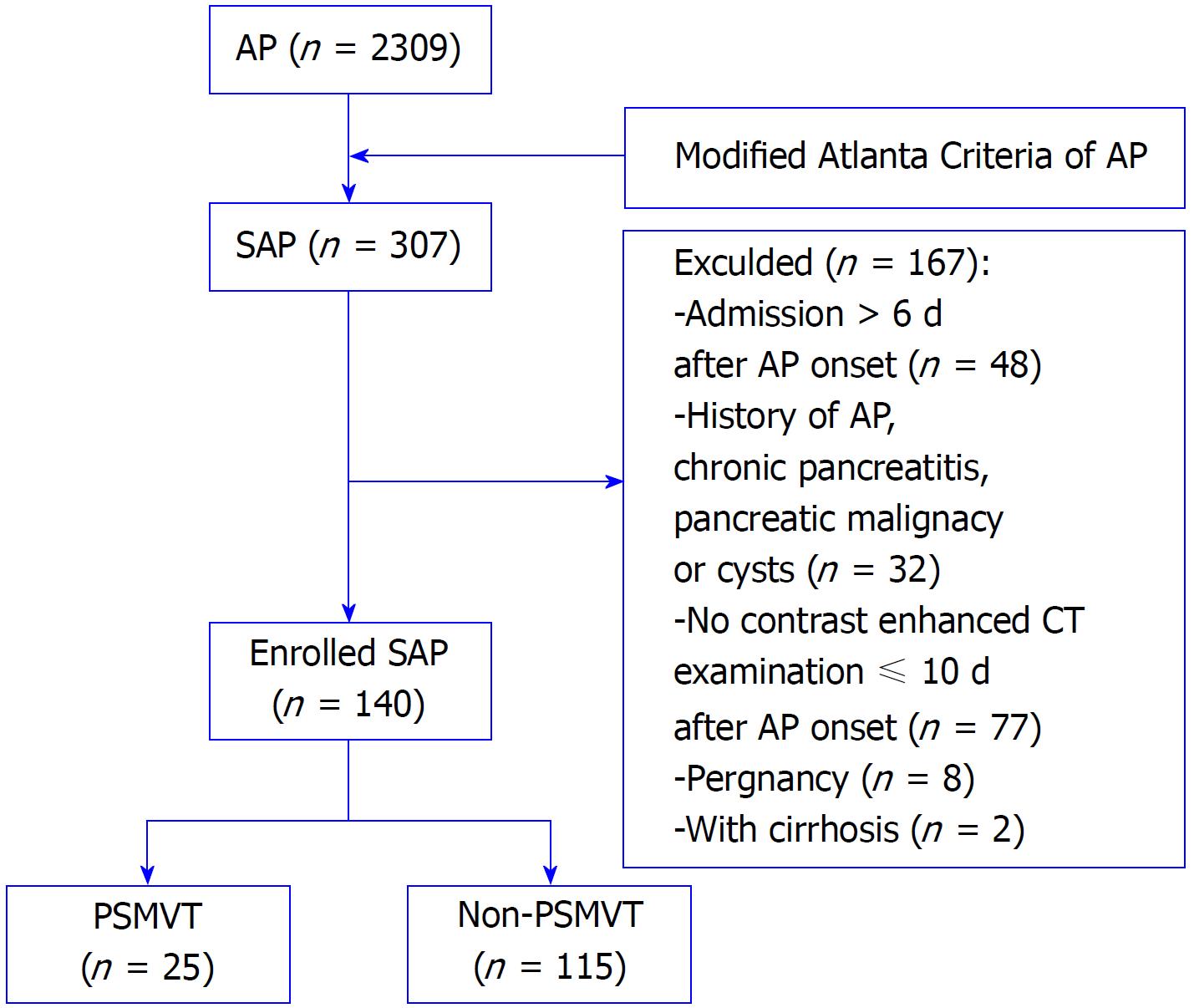
Figure 1 Flow chart of patients with or without portosplenomesenteric vein thrombosis secondary to severe acute pancreatitis in the early stage.
AP: Acute pancreatitis; SAP: Severe acute pancreatitis; CT: Computed tomography; PSMVT: Portosplenomesenteric vein thrombosis.
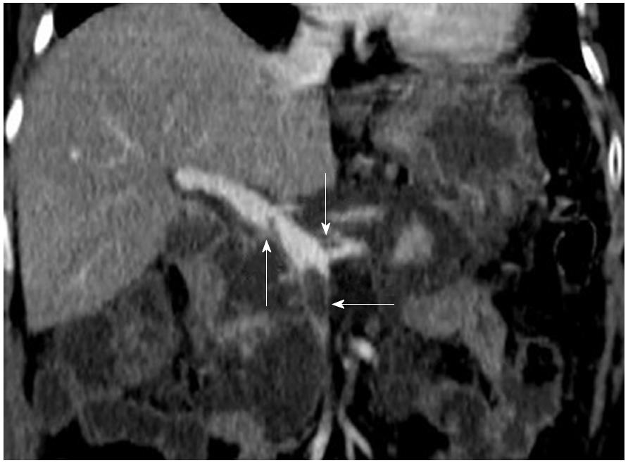
Figure 2 Portosplenomesenteric vein thrombosis.
Contrast-enhanced CT image in a 70-year-old female patient with severe acute biliary pancreatitis shows a filling defect within the lumen of the portal vein (up arrow), splenic vein (down arrow) and superior mesenteric vein (left arrow). CT: Computed tomography
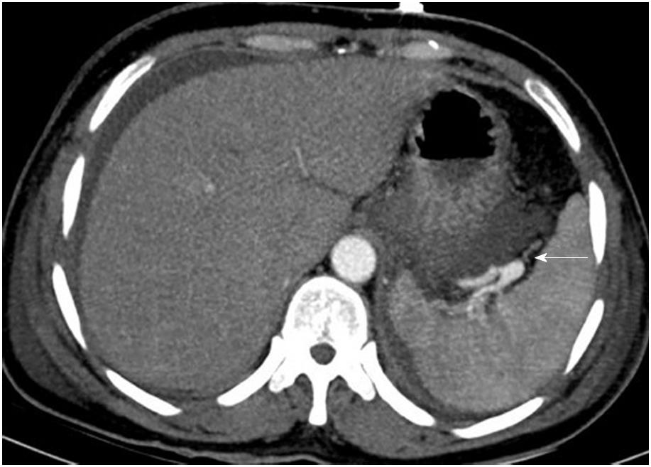
Figure 3 Pseudoaneurysm.
Axial contrast-enhanced CT image in a 42-year-old female patient with severe acute biliary pancreatitis shows a pseudoaneurysm of the splenic artery (arrow), and the patient was discharged without gastrointestinal hemorrhage or intra-abdominal hemorrhage.
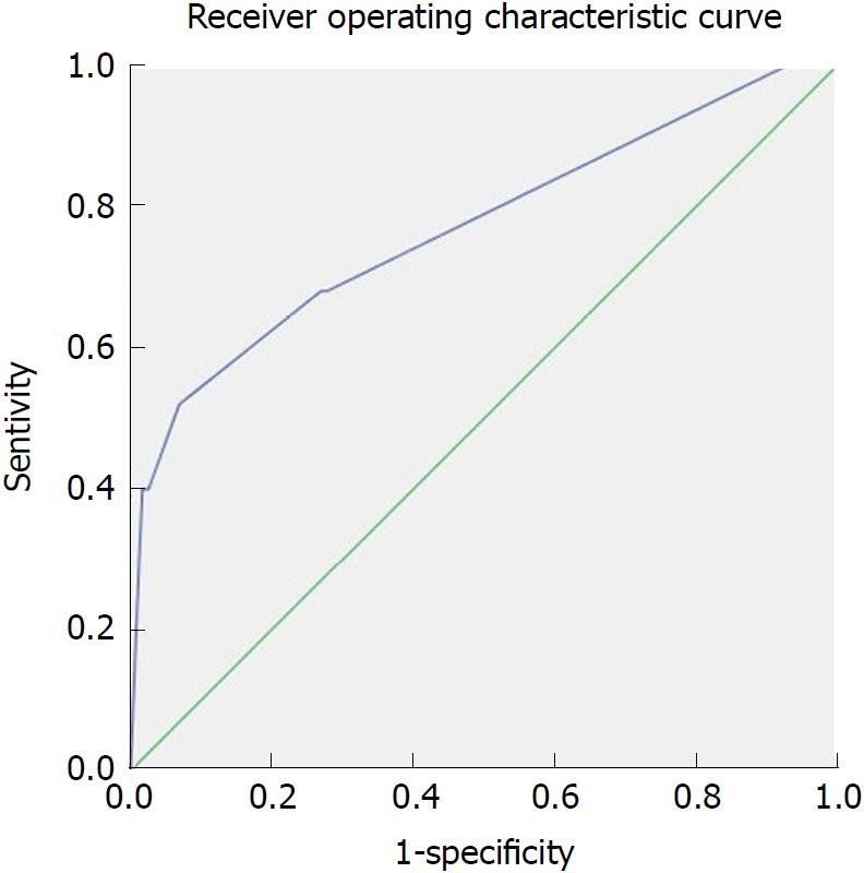
Figure 4 The receiver operating characteristic curve for Balthazar’s computed tomography severity index score in predicting PSMVT secondary to severe acute pancreatitis in the early stage.
- Citation: Ding L, Deng F, Yu C, He WH, Xia L, Zhou M, Huang X, Lei YP, Zhou XJ, Zhu Y, Lu NH. Portosplenomesenteric vein thrombosis in patients with early-stage severe acute pancreatitis. World J Gastroenterol 2018; 24(35): 4054-4060
- URL: https://www.wjgnet.com/1007-9327/full/v24/i35/4054.htm
- DOI: https://dx.doi.org/10.3748/wjg.v24.i35.4054


