Copyright
©The Author(s) 2018.
World J Gastroenterol. Jan 21, 2018; 24(3): 438-444
Published online Jan 21, 2018. doi: 10.3748/wjg.v24.i3.438
Published online Jan 21, 2018. doi: 10.3748/wjg.v24.i3.438
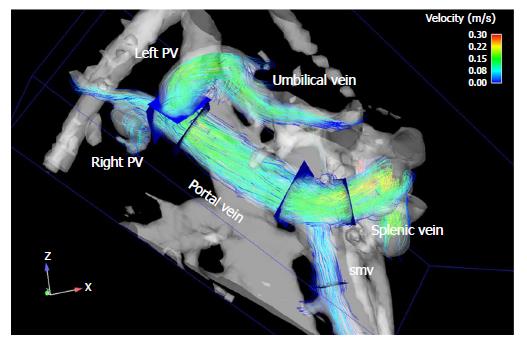
Figure 1 Time-resolved 3D MRI image shows prograde blood flow in the extrahepatic portal venous system and a recanalization of the umbilical vein with flow over the left branch of the intrahepatic portal vein.
PV: Portal vein; smv: Superior mesenteric vein.
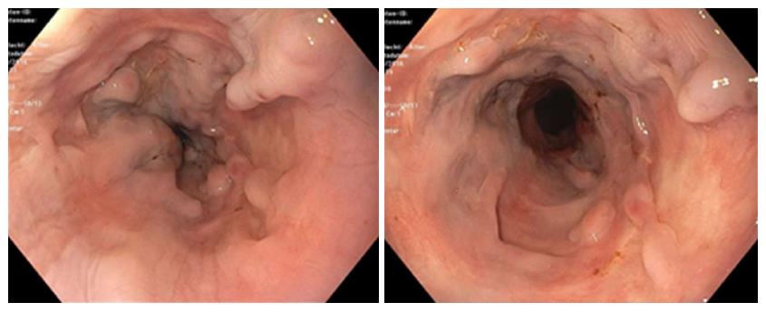
Figure 2 Distal aspect of the esophagus (12/2016).
Endoscopy shows scarring in the distal esophagus caused by sclerosing and banding, the old varices remain thrombosed.
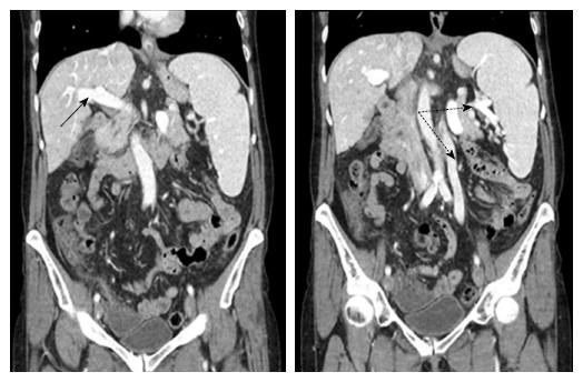
Figure 3 Computer tomographic angiography of the abdomen (09/2017).
These sections show the normal sized-cirrhotic liver with the portal vein draining blood in prograde direction (solid arrow). The spleen is considerably enlarged. There is a collateral blood flow originating from the splenic hilus via an enlarged ovarian vein to the pelvis (dashed arrows).
- Citation: Deibert P, Lazaro A, Stankovic Z, Schaffner D, Rössle M, Kreisel W. Beneficial long term effect of a phosphodiesterase-5-inhibitor in cirrhotic portal hypertension: A case report with 8 years follow-up. World J Gastroenterol 2018; 24(3): 438-444
- URL: https://www.wjgnet.com/1007-9327/full/v24/i3/438.htm
- DOI: https://dx.doi.org/10.3748/wjg.v24.i3.438


