Copyright
©The Author(s) 2018.
World J Gastroenterol. Jul 14, 2018; 24(26): 2893-2901
Published online Jul 14, 2018. doi: 10.3748/wjg.v24.i26.2893
Published online Jul 14, 2018. doi: 10.3748/wjg.v24.i26.2893
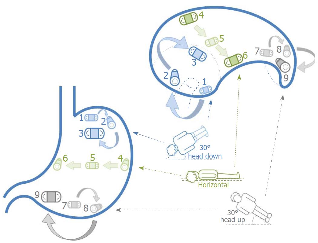
Figure 1 Schematic of the simple positional interchange technique.
Coronal views are illustrated on the left and transverse views (with the cranial end closest to the reader) on the right. Capsule movement is achieved by exploiting the effects of water flow from one gravity dependent area to another with patient positional change. Once the UGI capsule enters the stomach, the examination bed is tilted 30° head down (depicted in blue) and patients lie supine (position 1), on their left lateral (position 2) and then prone (position 3). The bed is returned to the horizontal plane (depicted in green) and patients lie on their left lateral (position 4), supine (position 5) and then right lateral (position 6). The bed is finally adjusted to 30° head up (depicted in grey) and patients lie supine (position 7), on their left lateral (position 8) and then prone (position 9). UGI: Upper gastrointestinal.
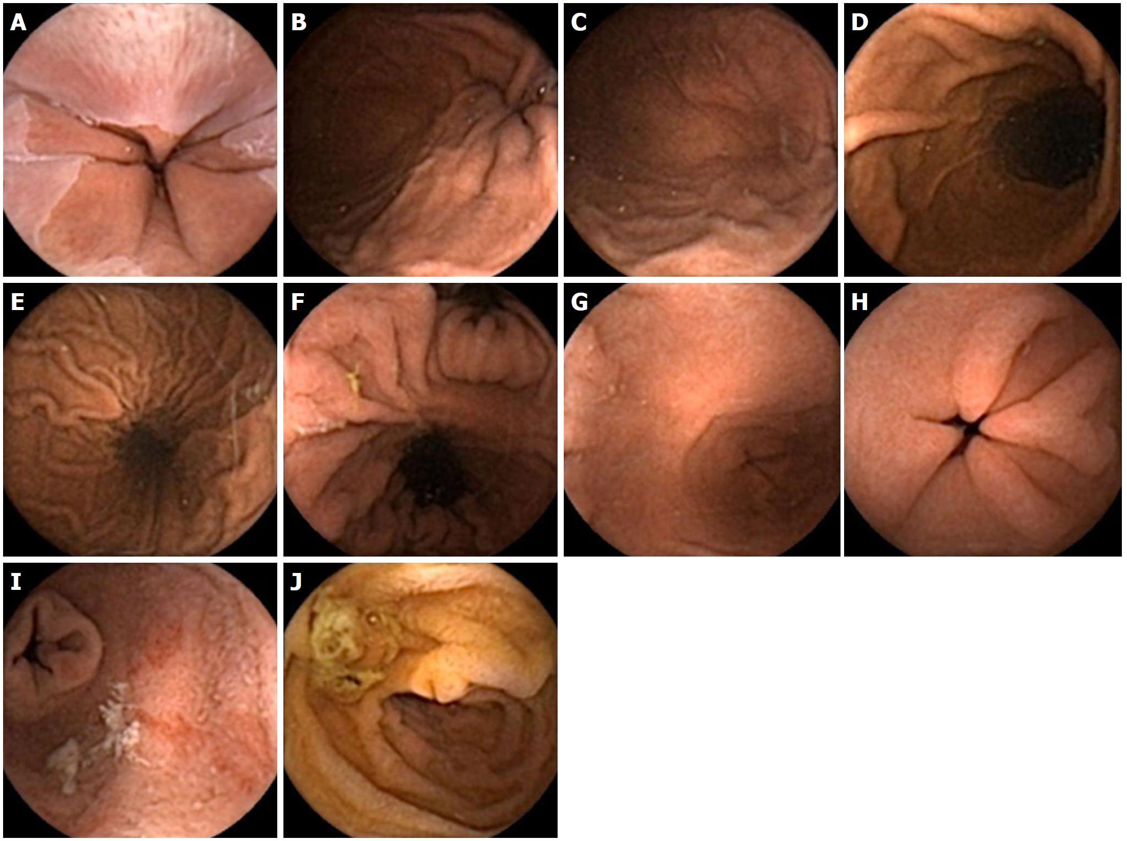
Figure 2 Normal views of the upper gastrointestinal tract seen with the upper gastrointestinal capsule.
A: Gastroesophageal junction; B: Cardia; C: Fundus; D: Greater curvature; E: Lesser curvature; F: Incisura angularis; G: Antrum; H: Pylorus; I: First part of duodenum (retrograde view); J: Second part of duodenum (ampulla also seen).
Figure 3 Suboptimal views in the fundus.
A: Mucus; B: Bubbles; C: Insufficient distension.
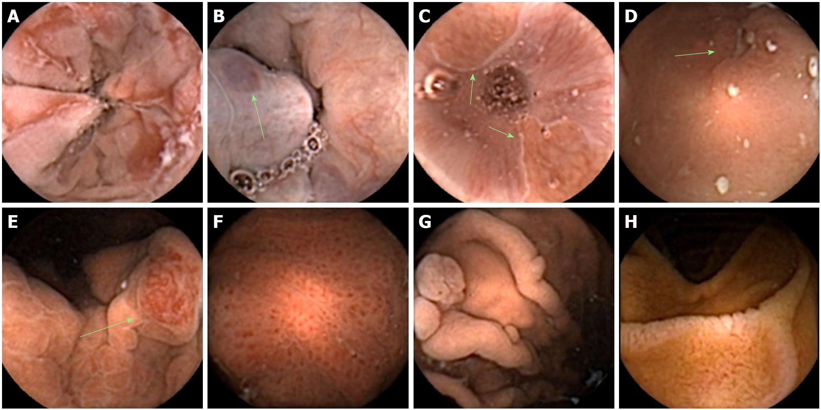
Figure 4 Pathology detected by upper gastrointestinal capsule.
A: Erosive esophagitis; B: Oesophageal varices; C: Barrett’s oesophagus; D: Gastric ulcer; E: Gastric angioectasia; F: Portal hypertensive gastropathy; G: Benign cystic fundic gland polyps; H: Coeliac disease.
- Citation: Ching HL, Healy A, Thurston V, Hale MF, Sidhu R, McAlindon ME. Upper gastrointestinal tract capsule endoscopy using a nurse-led protocol: First reported experience. World J Gastroenterol 2018; 24(26): 2893-2901
- URL: https://www.wjgnet.com/1007-9327/full/v24/i26/2893.htm
- DOI: https://dx.doi.org/10.3748/wjg.v24.i26.2893


