Copyright
©The Author(s) 2018.
World J Gastroenterol. Jul 14, 2018; 24(26): 2893-2901
Published online Jul 14, 2018. doi: 10.3748/wjg.v24.i26.2893
Published online Jul 14, 2018. doi: 10.3748/wjg.v24.i26.2893
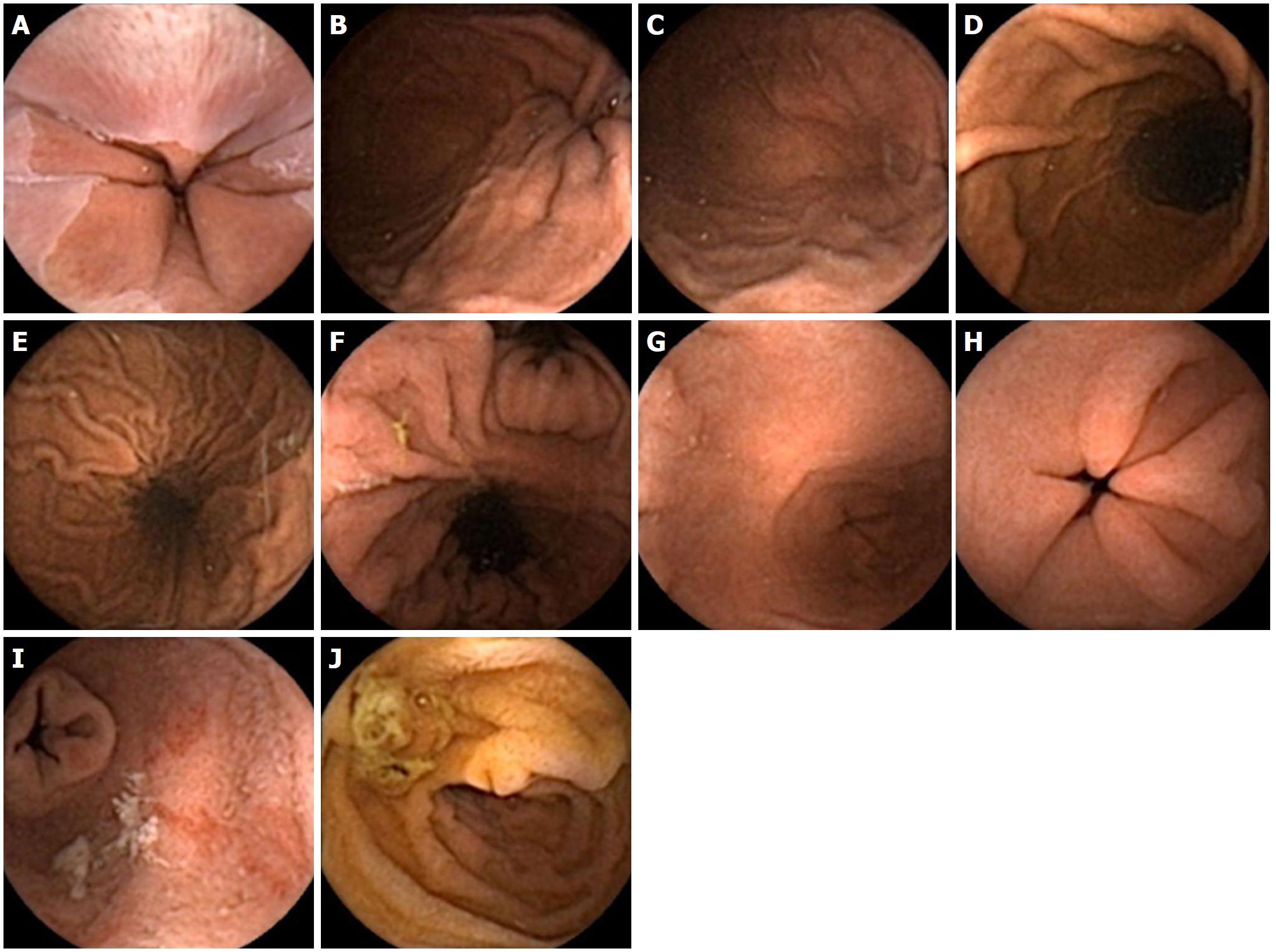
Figure 2 Normal views of the upper gastrointestinal tract seen with the upper gastrointestinal capsule.
A: Gastroesophageal junction; B: Cardia; C: Fundus; D: Greater curvature; E: Lesser curvature; F: Incisura angularis; G: Antrum; H: Pylorus; I: First part of duodenum (retrograde view); J: Second part of duodenum (ampulla also seen).
- Citation: Ching HL, Healy A, Thurston V, Hale MF, Sidhu R, McAlindon ME. Upper gastrointestinal tract capsule endoscopy using a nurse-led protocol: First reported experience. World J Gastroenterol 2018; 24(26): 2893-2901
- URL: https://www.wjgnet.com/1007-9327/full/v24/i26/2893.htm
- DOI: https://dx.doi.org/10.3748/wjg.v24.i26.2893


