Copyright
©The Author(s) 2018.
World J Gastroenterol. Apr 7, 2018; 24(13): 1410-1418
Published online Apr 7, 2018. doi: 10.3748/wjg.v24.i13.1410
Published online Apr 7, 2018. doi: 10.3748/wjg.v24.i13.1410
Figure 1 Mini-laparoscopy of a patient with acute liver failure due to newly diagnosed autoimmune hepatitis exemplarily showing the right liver lobe with diffuse capsular swelling, regenerative nodules, and rounded lower margin (upper panel) (A), and liver histology of the same patient with AIH-induced ALF (B).
Left panel demonstrating severe inflammation with interface hepatitis (original magnification 200 ×) and the right panel with higher magnification (400 ×) revealing numerous plasma cells extending from the portal tract into the adjacent parenchyma (lower panel).
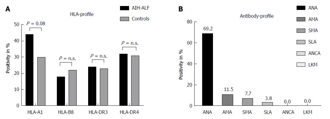
Figure 2 HLA-profile of the study population (n = 34) investigating HLA-A1, -B8, -DR3, and -DR4 status (A), and antibody-profile of the study population (n = 52) demonstrating positivity for ANA-, AMA-, SMA-, SLA-, ANCA-, and LKM-titers (B).
ANA: Anti-nuclear; SMA: Anti-smooth muscle; LKM: Anti-liver kidney microsomal.
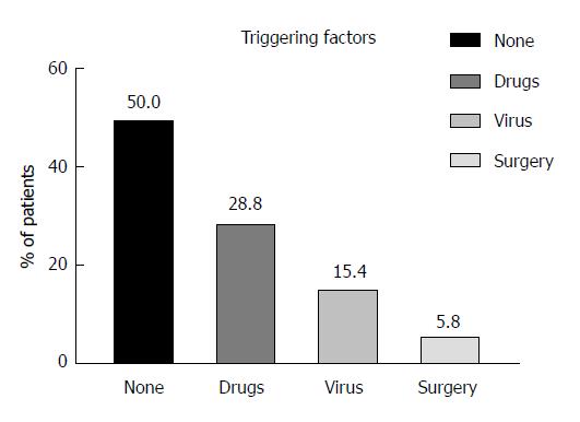
Figure 3 Potential triggering factors for acute liver failure in patients with their first manifestation of autoimmune hepatitis (n = 52).
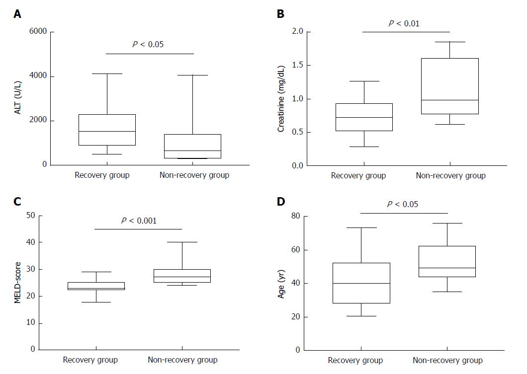
Figure 4 Higher age, creatinine, and MELD-score were associated with lethal outcome.
A: Median alanine-aminotransferase (ALT)-values of patients with recovery as compared to the non-recovery group, P < 0.05; B: Median serum creatinine levels of patients with recovery and non-recovery, P < 0.01; C: Median labMELD-score of patients with recovery and non-recovery, P < 0.001; D: Median age of patients with recovery and non-recovery, P < 0.05.
- Citation: Buechter M, Manka P, Heinemann FM, Lindemann M, Baba HA, Schlattjan M, Canbay A, Gerken G, Kahraman A. Potential triggering factors of acute liver failure as a first manifestation of autoimmune hepatitis-a single center experience of 52 adult patients. World J Gastroenterol 2018; 24(13): 1410-1418
- URL: https://www.wjgnet.com/1007-9327/full/v24/i13/1410.htm
- DOI: https://dx.doi.org/10.3748/wjg.v24.i13.1410


