Copyright
©The Author(s) 2017.
World J Gastroenterol. Feb 21, 2017; 23(7): 1171-1179
Published online Feb 21, 2017. doi: 10.3748/wjg.v23.i7.1171
Published online Feb 21, 2017. doi: 10.3748/wjg.v23.i7.1171
Figure 1 Thymoquinone affects the viability of LoVo colon cancer cells.
To explore the effect of thymoquinone (TQ) on the viability of human LoVo colon cancer cells, we first treated LoVo cells with various concentrations (2.5, 5, 7.5, 10 and 20 μmol/L) of TQ for 24 h and subsequently measured cell viability by MTT assay. The results showed a significant reduction of cell viability of approximately 60% following treatment (20 μmol/L) for 24 h. Each value represents the mean ± SE. aP < 0.05; bP < 0.01.
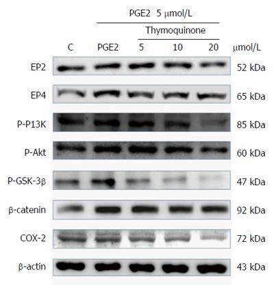
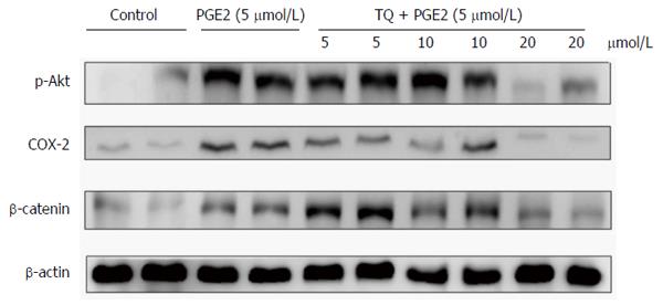
Figure 2 Thymoquinone inhibits the expression of PI3K, Akt, p-GSK3β, β-catenin and COX-2 proteins in LoVo cells.
LoVo cells were cultured in medium that was treated with TQ (5, 10 and 20 μmol/L) for 24 h; PGE2 (5 μmol/L) was added for the last 6 h. Following this, the cells were immunoblotted with the indicated antibodies. The cells were lysed, and the extracts were separated by 10% SDS-PAGE, transferred to PVDF membranes and immunoblotted with antibodies against p-PI3K, p-Akt, p-GSK3β, β-catenin and COX-2.
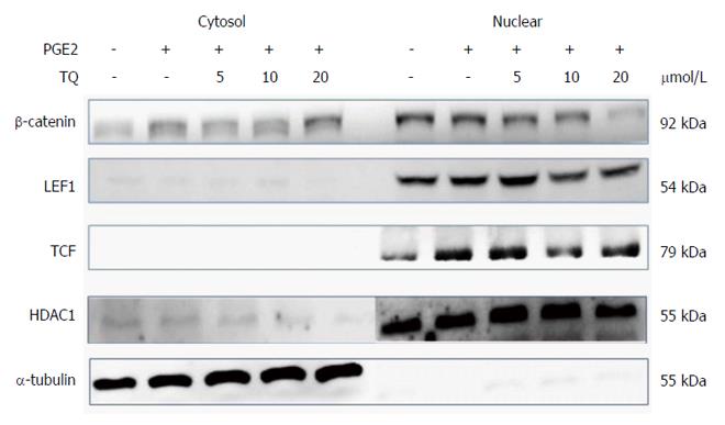
Figure 3 Thymoquinone inhibits β-catenin nuclear translocation in the LoVo cancer cell line.
Nuclear isolation showed a decrease in β-catenin translocation into the nucleus. The cofactors LEF-1 and TCF-4 decreased in the nucleus following TQ treatment in a dose-dependent manner. TQ: Thymoquinone.
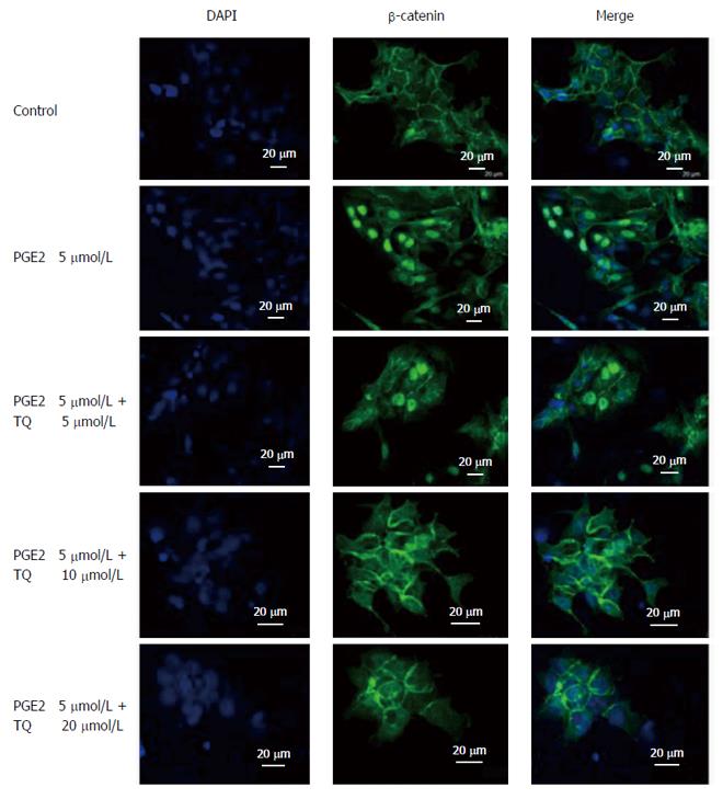
Figure 4 Thymoquinone treatment inhibits β-catenin translocation into LoVo cell nuclei.
An immunofluorescence assay was performed on LoVo cells using a β-catenin primary antibody and a secondary antibody (1:250) producing green fluorescence; DAPI (blue fluorescence) was included to stain cell nuclei. Merged β-catenin and DAPI (green and blue, respectively) signals are shown. The indicated treatments were assessed. PGE2: Prostaglandin E2; TQ: Thymoquinone.
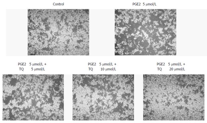
Figure 5 Thymoquinone efficiently inhibits LoVo cell migration.
LoVo cells were pretreated with increasing dosages (5, 10, and 20 μmol/L) of TQ for 24 h. A Boyden chamber migration assay was performed to assess cell migration ability. The responses to different treatments were analyzed via microscopy. PGE2: Prostaglandin E2; TQ: Thymoquinone.
Figure 6 Impact of thymoquinone and/or prostaglandin E2 administration on tumor growth in nude mice xenografts.
Tumor tissues were harvested from nude mice and lysed. Protein content was quantified and analyzed by immunoblotting. p-Akt, β-catenin and COX-2 levels in LoVo cells were detected. PGE2: Prostaglandin E2; TQ: Thymoquinone.
- Citation: Hsu HH, Chen MC, Day CH, Lin YM, Li SY, Tu CC, Padma VV, Shih HN, Kuo WW, Huang CY. Thymoquinone suppresses migration of LoVo human colon cancer cells by reducing prostaglandin E2 induced COX-2 activation. World J Gastroenterol 2017; 23(7): 1171-1179
- URL: https://www.wjgnet.com/1007-9327/full/v23/i7/1171.htm
- DOI: https://dx.doi.org/10.3748/wjg.v23.i7.1171


