Copyright
©The Author(s) 2017.
World J Gastroenterol. Jul 21, 2017; 23(27): 5034-5040
Published online Jul 21, 2017. doi: 10.3748/wjg.v23.i27.5034
Published online Jul 21, 2017. doi: 10.3748/wjg.v23.i27.5034
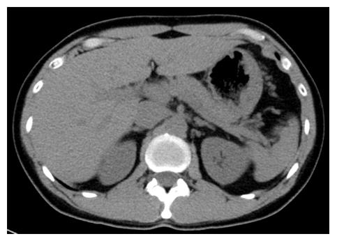
Figure 1 Abdominal computed tomography scans did not show any morphological changes on admission.
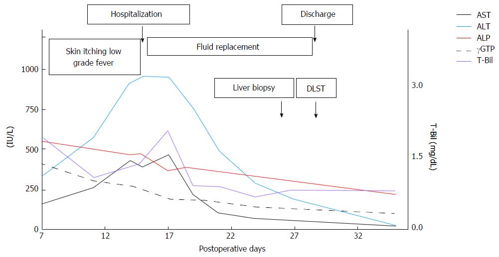
Figure 2 Aspartate aminotransferase, alanine aminotransferase, alkaline phosphatase, total bilirubin, and gamma-glutamyl transpeptidase levels in the patient at presentation and in response to treatment.
AST: Aspartate aminotransferase; ALT: Alanine aminotransferase; ALP: Alkaline phosphatase; γGTP: Gamma-glutamyl transpeptidase; T-Bil: Total bilirubin; DLST: Drug lymphocyte stimulation test.
Figure 3 Micrographs of a liver biopsy specimen.
A: Fibrosis around the central vein area and the fibrous expansion of the portal region were not detected (Masson staining × 40); B: Swelling of the parenchymal cells and a mild infiltration of inflammatory cells around the central vein area were evident (green arrows). Destruction of the bile duct had not occurred (hematoxylin-eosin staining × 40); C: Bile plugs were found in multiple sites within the hepatocytes and sinusoids (green arrows). (hematoxylin-eosin staining × 200). CV: Central vein; PV: Portal vein.
- Citation: Yoshikawa K, Kawashima R, Hirose Y, Shibata K, Akasu T, Hagiwara N, Yokota T, Imai N, Iwaku A, Kobayashi G, Kobayashi H, Kinoshita A, Fushiya N, Kijima H, Koike K, Saruta M. Liver injury after aluminum potassium sulfate and tannic acid treatment of hemorrhoids. World J Gastroenterol 2017; 23(27): 5034-5040
- URL: https://www.wjgnet.com/1007-9327/full/v23/i27/5034.htm
- DOI: https://dx.doi.org/10.3748/wjg.v23.i27.5034


