Copyright
©The Author(s) 2017.
World J Gastroenterol. Jul 21, 2017; 23(27): 4950-4957
Published online Jul 21, 2017. doi: 10.3748/wjg.v23.i27.4950
Published online Jul 21, 2017. doi: 10.3748/wjg.v23.i27.4950
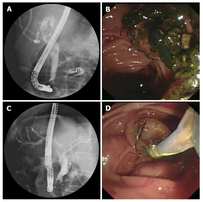
Figure 1 Large stones in the common bile duct were cracked by extracorporeal shock wave lithotripsy (A) and cleared by following endoscopic retrograde cholangiopancreatography (B and C), common bile duct strictures were dilated using a balloon, and passage dilating catheters were used to retrieve the stones (D).
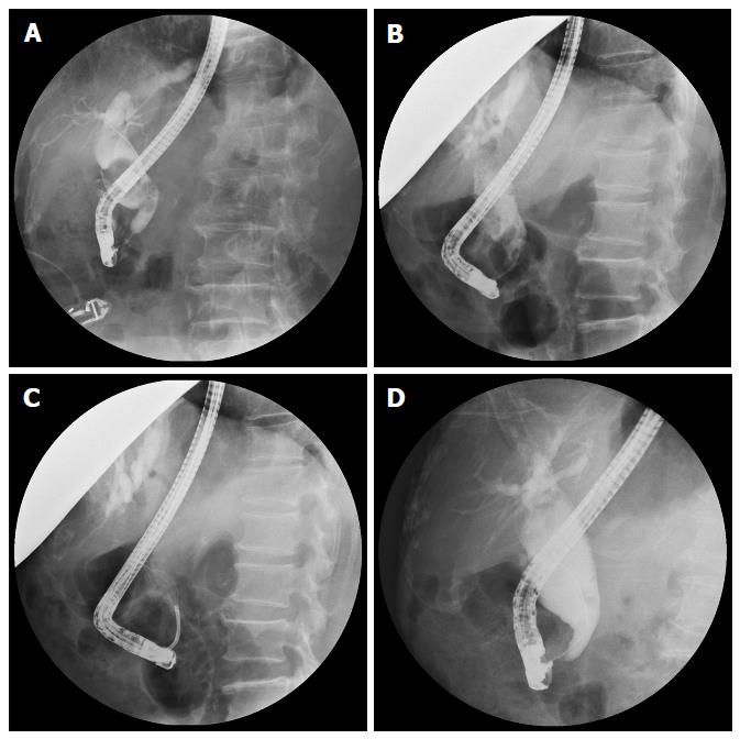
Figure 2 Common bile duct stone clearance was assessed after each endoscopic retrograde cholangiopancreatography session using procedure reports, plain films, endoscopic retrograde cholangiopancreatography films and/or abdominal magnetic resonance cholangiopancreatography.
A: Pre-ESWL large common bile duct stones were very large; B: Post-ESWL reduction in diameter of CBD stones; C: Stones were tracked by a basket during the following ERCP; D: CBD was cleared successfully. ESWT: Extracorporeal shockwave lithotripsy; ERCP: Endoscopic retrograde cholangiopancreatography; CBD: Common bile duct.
- Citation: Tao T, Zhang M, Zhang QJ, Li L, Li T, Zhu X, Li MD, Li GH, Sun SX. Outcome of a session of extracorporeal shock wave lithotripsy before endoscopic retrograde cholangiopancreatography for problematic and large common bile duct stones. World J Gastroenterol 2017; 23(27): 4950-4957
- URL: https://www.wjgnet.com/1007-9327/full/v23/i27/4950.htm
- DOI: https://dx.doi.org/10.3748/wjg.v23.i27.4950


