Copyright
©The Author(s) 2017.
World J Gastroenterol. Jul 14, 2017; 23(26): 4712-4723
Published online Jul 14, 2017. doi: 10.3748/wjg.v23.i26.4712
Published online Jul 14, 2017. doi: 10.3748/wjg.v23.i26.4712
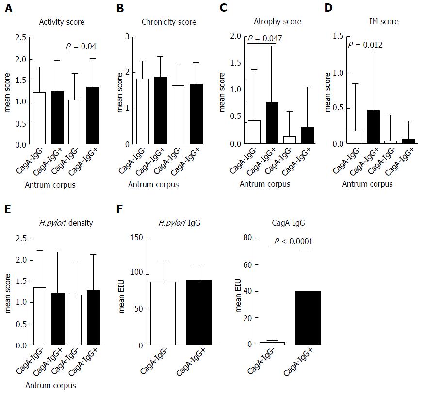
Figure 1 Difference in Sydney score and Helicobacter pylori seropositivity in patients with and without anti-CagA-IgG.
Mean histological scores ± SD. A: Activity; B: Chronicity, C: Atrophy; D: Intestinal metaplasia are shown; E: Anti-Helicobacter pylori IgG; F: anti-CagA-IgG titer were evaluated using ELISA; Statistical analyses were performed using Mann-Whitney test.
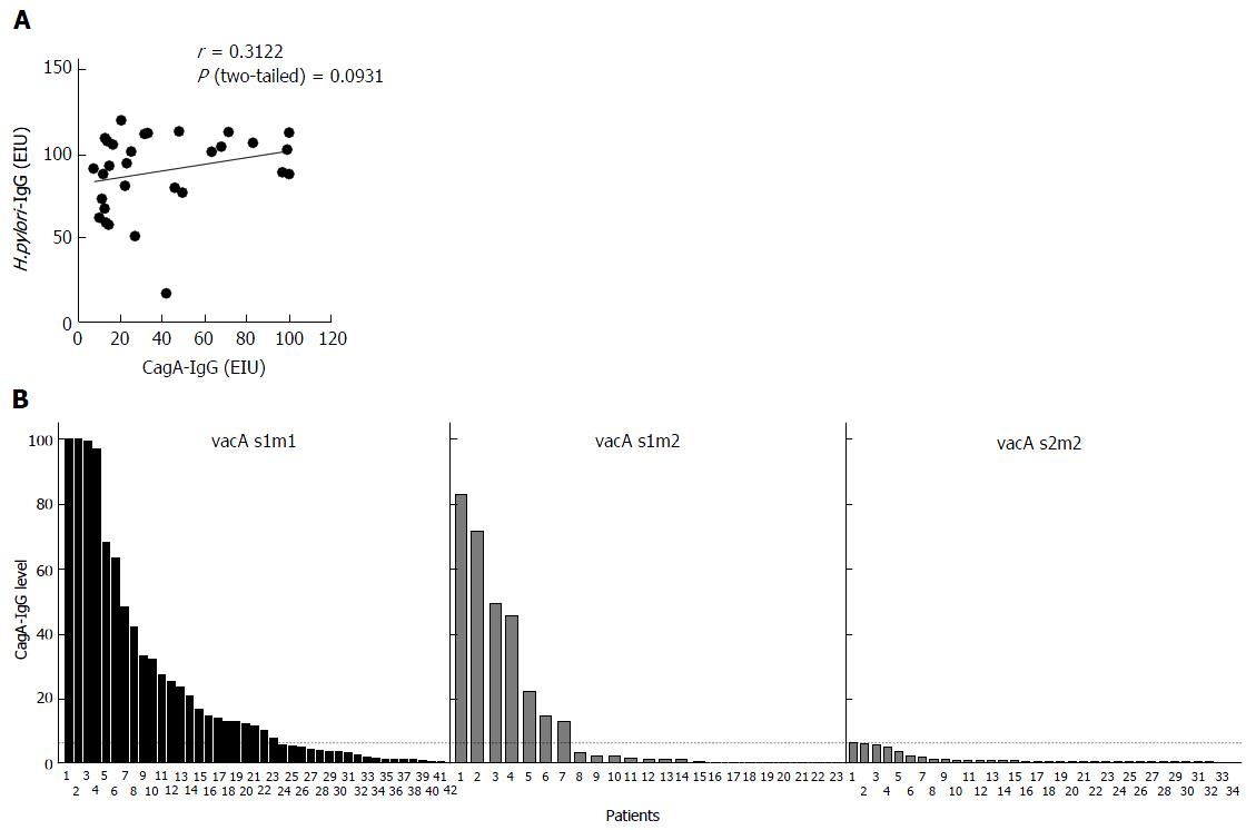
Figure 2 Correlation between anti-CagA-IgG and anti-Helicobacter pylori IgG and vacA s/m polymorphisms.
A: Quantitative values for anti-Helicobacter pylori IgG and anti-CagA-IgG were correlated in patients with positive serology (n = 30) using Pearson’s test; B: Patients with available data to vacA s1/2m1/2 polymorphisms were divided in subgroups dependent on s/m subtype. Anti-CagA-IgG values were sorted in increasing order. Dotted line shows the cut-off for seropositivity of the test (6.25 U/mL).
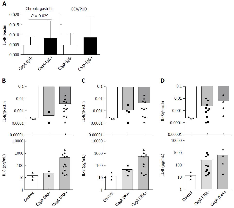
Figure 3 Inflammatory potential based on CagA-IgG in gastric mucosa ex vivo and in vitro using AGS cells.
A: IL-8 mRNA expression was evaluated in antrum mucosa from patients with chronic gastritis [CG, AG, IM, without peptic ulcer disease (PUD) and gastric cancer (GC)] with (n = 23) and without CagA seropositivity (n = 48); B: IL-8 expression in antrum mucosa from patients with PUD and gastric cancer (n = 4 for CagA-IgG+ and n = 6 CagA-IgG-); C: IL-8 mRNA; D: IL-8 protein expression in supernatant are shown for subgroups dependent on cagA, cagA mRNA and anti-CagA-IgG status in vitro using co-culturing of Helicobacter pylori strains from patients with AGS cells.
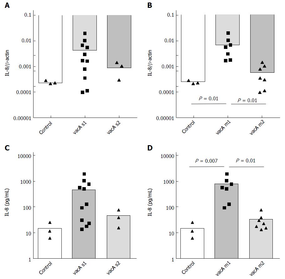
Figure 4 Inflammatory potential of Helicobacter pylori strains in relation to vacA polymorphism.
Helicobacter pylori strains from patients were co-cultivated with AGS cell and A-B: IL-8 mRNA; C-D: IL-8 expression in supernatant were measured using qPCR and ELISA.
- Citation: Link A, Langner C, Schirrmeister W, Habendorf W, Weigt J, Venerito M, Tammer I, Schlüter D, Schlaermann P, Meyer TF, Wex T, Malfertheiner P. Helicobacter pylori vacA genotype is a predominant determinant of immune response to Helicobacter pylori CagA. World J Gastroenterol 2017; 23(26): 4712-4723
- URL: https://www.wjgnet.com/1007-9327/full/v23/i26/4712.htm
- DOI: https://dx.doi.org/10.3748/wjg.v23.i26.4712


