Copyright
©The Author(s) 2017.
World J Gastroenterol. Jun 21, 2017; 23(23): 4303-4310
Published online Jun 21, 2017. doi: 10.3748/wjg.v23.i23.4303
Published online Jun 21, 2017. doi: 10.3748/wjg.v23.i23.4303
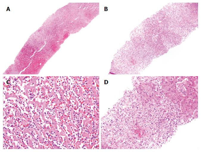
Figure 1 Mass and submassive liver necrosis.
A: Low power magnification showing complete hepatocellular necrosis in a core biopsy (× 40); B: Medium power magnification showing a liver biopsy with perivenular predominant hepatocellular necrosis and intermixed viable hepatocytes located around the portal tract (× 40); C: High magnification of image A showing hepatocellular necrosis and profound inflammatory infiltrate in which the architecture of hepatocyte is preserved (× 200); D: High magnification of image B reveals the collapse of parenchyma that is replaced by necroinflammatory infiltrate in comparison to the adjacent viable hepatocytes in the right upper corner (× 200).
- Citation: Ndekwe P, Ghabril MS, Zang Y, Mann SA, Cummings OW, Lin J. Substantial hepatic necrosis is prognostic in fulminant liver failure. World J Gastroenterol 2017; 23(23): 4303-4310
- URL: https://www.wjgnet.com/1007-9327/full/v23/i23/4303.htm
- DOI: https://dx.doi.org/10.3748/wjg.v23.i23.4303


