Copyright
©The Author(s) 2017.
World J Gastroenterol. May 14, 2017; 23(18): 3338-3348
Published online May 14, 2017. doi: 10.3748/wjg.v23.i18.3338
Published online May 14, 2017. doi: 10.3748/wjg.v23.i18.3338
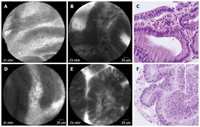
Figure 1 In vivo endoscopic ultrasound guided needle based confocal laser endomicroscopy, ex vivo confocal laser endomicroscopy, and histopathology of intraductal papillary mucinous neoplasms.
Panels A, B and C are from subject 1 (gastric subtype with high grade dysplasia). Panels D, E, and F are from subject 2 (intestinal subtype with high grade dysplasia). Complete “fingerlike” papillae are observed in both in vivo and ex vivo CLE. The vascular core in ex vivo CLE imaging is better defined. Histopathology (panels C, F): 40 × magnification; HE stain.
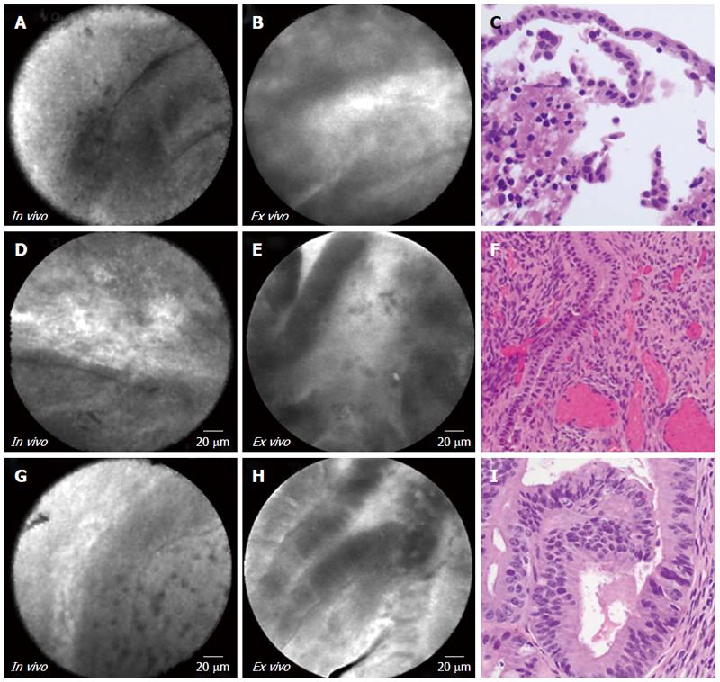
Figure 2 In vivo endoscopic ultrasound guided needle based confocal laser endomicroscopy, ex vivo confocal laser endomicroscopy, and histopathology of mucinous cystic neoplasms.
Panels A, B, and C are from subject 3 (low grade). Panels D, E, and F are from subject 4 (low grade). Panels G, H, and I are from subject 5 (low to moderate grade). Epithelial bands with incomplete papillary formation are observed in CLE. The in vivo CLE demonstrates horizon like bands where ex vivo CLE demonstrates better defined epithelial bands. Corresponding histopathology (panel C, × 40 and panel F, × 20) show low grade dysplasia and panel I (× 40) reveals moderate grade dysplasia. CLE: Confocal laser endomicroscopy.
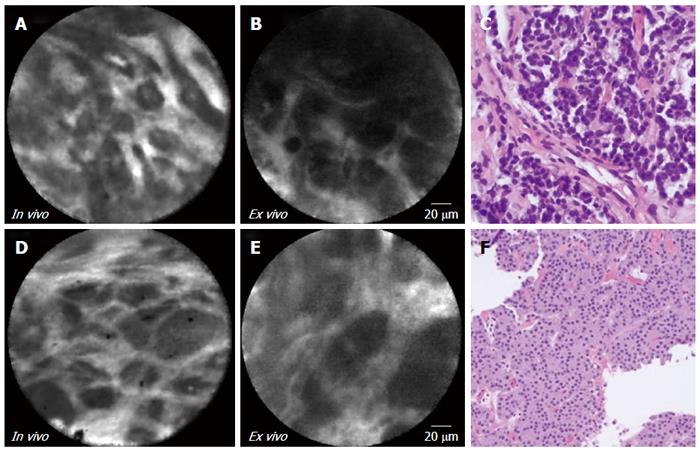
Figure 3 In vivo endoscopic ultrasound guided needle based confocal laser endomicroscopy, ex vivo confocal laser endomicroscopy, and histopathology of cystic neuroendocrine tumor.
Panels A, B, and C are from subject 6. Panels D, E, and F are from subject 7. Circumscribed clusters of cells in a trabecular growth pattern separated by vascular or fibrous cords are observed on confocal laser endomicroscopy examination. Histopathology (panels C, × 40; panel F, × 20) revealed characteristic uniform tumor cells arranged in cords or trabecular fashion.
Figure 4 In vivo endoscopic ultrasound guided needle based confocal laser endomicroscopy, ex vivo confocal laser endomicroscopy, and histopathology of serous cystadenoma.
Confocal laser endomicroscopy images, panels A (in vivo) and B (ex vivo) depict “fern pattern” of vascularity (subject 8). Histopathology (panel C; HE, × 40) reveals cuboidal to flat epithelial cells with clear cytoplasm lining some cystic spaces.
Figure 5 In vivo endoscopic ultrasound guided needle based confocal laser endomicroscopy, ex vivo confocal laser endomicroscopy, and histopathology of epidermoid cyst of intra pancreatic accessory spleen.
Confocal laser endomicroscopy images, panels A (in vivo) and B (ex vivo) reveal underlying splenic tissue (panels C, red pulp). Histopathology shows thin epithelial layer (squamous) with underlying ectopic splenic tissue (HE, × 40).
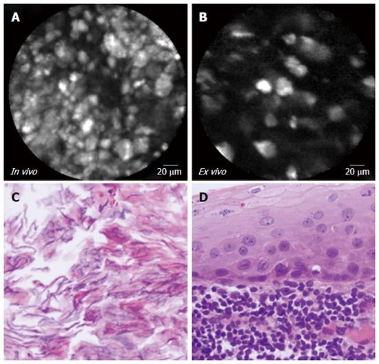
Figure 6 In vivo endoscopic ultrasound guided needle based confocal laser endomicroscopy, ex vivo confocal laser endomicroscopy, and histopathology of lymphoepithelial cyst.
Confocal laser endomicroscopy images, A (in vivo) and B (ex vivo) reveal clusters of bright particles representing keratin flakes. Macroscopically the lesion was filled with yellowish pasty material which by microscopy (panel C) demonstrated keratin flakes. The cyst was lined by squamous epithelium surrounded by abundant lymphoid tissue (panel D; HE, × 40).
- Citation: Krishna SG, Modi RM, Kamboj AK, Swanson BJ, Hart PA, Dillhoff ME, Manilchuk A, Schmidt CR, Conwell DL. In vivo and ex vivo confocal endomicroscopy of pancreatic cystic lesions: A prospective study. World J Gastroenterol 2017; 23(18): 3338-3348
- URL: https://www.wjgnet.com/1007-9327/full/v23/i18/3338.htm
- DOI: https://dx.doi.org/10.3748/wjg.v23.i18.3338


