
















©The Author(s) 2017.
World J Gastroenterol. May 14, 2017; 23(18): 3338-3348
Published online May 14, 2017. doi: 10.3748/wjg.v23.i18.3338
Published online May 14, 2017. doi: 10.3748/wjg.v23.i18.3338
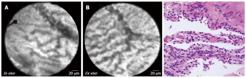
Figure 4 In vivo endoscopic ultrasound guided needle based confocal laser endomicroscopy, ex vivo confocal laser endomicroscopy, and histopathology of serous cystadenoma.
Confocal laser endomicroscopy images, panels A (in vivo) and B (ex vivo) depict “fern pattern” of vascularity (subject 8). Histopathology (panel C; HE, × 40) reveals cuboidal to flat epithelial cells with clear cytoplasm lining some cystic spaces.
- Citation: Krishna SG, Modi RM, Kamboj AK, Swanson BJ, Hart PA, Dillhoff ME, Manilchuk A, Schmidt CR, Conwell DL. In vivo and ex vivo confocal endomicroscopy of pancreatic cystic lesions: A prospective study. World J Gastroenterol 2017; 23(18): 3338-3348
- URL: https://www.wjgnet.com/1007-9327/full/v23/i18/3338.htm
- DOI: https://dx.doi.org/10.3748/wjg.v23.i18.3338