Copyright
©The Author(s) 2017.
World J Gastroenterol. May 7, 2017; 23(17): 3053-3065
Published online May 7, 2017. doi: 10.3748/wjg.v23.i17.3053
Published online May 7, 2017. doi: 10.3748/wjg.v23.i17.3053
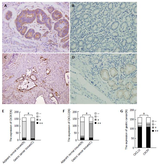
Figure 1 CXCR7 and CXCL12 expression in non-tumoral tissues and gastric cancer.
A: Gastric cancer tissue shows strong expression of CXCR7; B: Non-tumoral gastric tissue shows negative expression of CXCR7; C: Gastric cancer tissue shows strong expression of CXCL12; D: Non-tumoral gastric tissue shows weak expression of CXCL12; E: CXCR7 expression in cancer tissues was significantly higher than that in normal tissues; F: CXCL12 expression in cancer tissues was significantly higher than that in normal tissues; G: The expression of CXCR7 and CXCL12 was correlative in gastric cancer tissues. aP < 0.05, between the two groups.
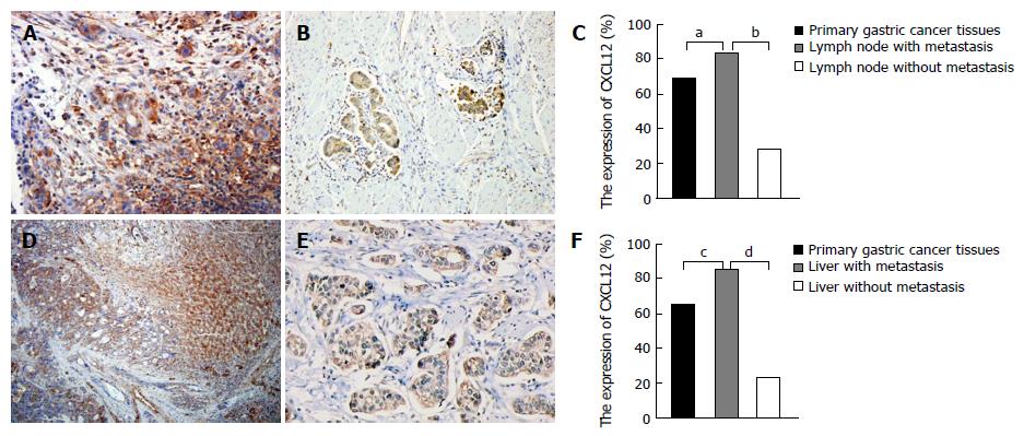
Figure 2 CXCL12 expression in lymph node, liver and primary gastric cancer tissues.
A: Metastatic lymph node shows strong expression of CXCL12; B: Primary gastric tissue shows strong expression of CXCL12; C: CXCL12 expression in lymph nodes with metastasis was significantly higher than that in primary gastric tissue and lymph node with no metastasis; D: Liver metastasis shows strong expression of CXCL12; E: Primary gastric tissue shows strong expression of CXCL12; F: CXCL12 expression in liver metastasis was significantly higher than that in primary gastric tissue and liver tissue with no metastasis. a-dP < 0.05, between the two groups.
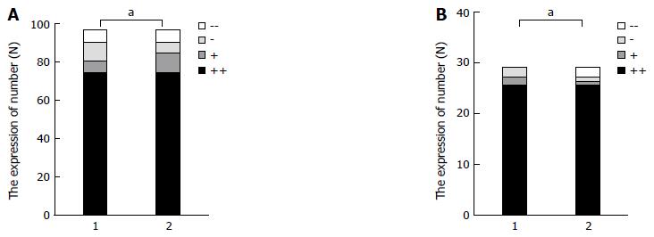
Figure 3 The relation of CXCL12 and CXCR7 expression.
A: Correlation analysis of CXCL12 expression in lymph nodes and CXCR7 expression in gastric cancer; B: Correlation analysis of CXCL12 expression in liver tissue and CXCR7 expression in gastric cancer. aP < 0.05, between the two groups. 1: The express of CXCL12 in lymph nodes with metastasis; 2: The express of CXCR7 in primary gastric cancer tissues
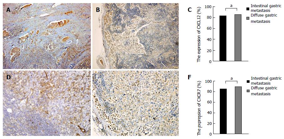
Figure 4 CXCL12/CXCR7 expression in intestinal-type gastric cancer and diffuse-type gastric cancer.
A: CXCL12 expression in lymph node metastasis of intestinal-type gastric cancer; B: CXCL12 expression in lymph node metastasis of diffuse-type gastric cancer; C: CXCL12 expression showed no significant difference between lymph node metastasis of intestinal-type and diffuse-type gastric cancer; D: CXCR7 expression in lymph node metastasis of intestinal-type gastric cancer; E: CXCR7 expression in lymph node metastasis of diffuse-type gastric cancer; F: CXCR7 expression showed no significant difference between lymph node metastasis of intestinal-type and diffuse-type gastric cancer. aP > 0.05.
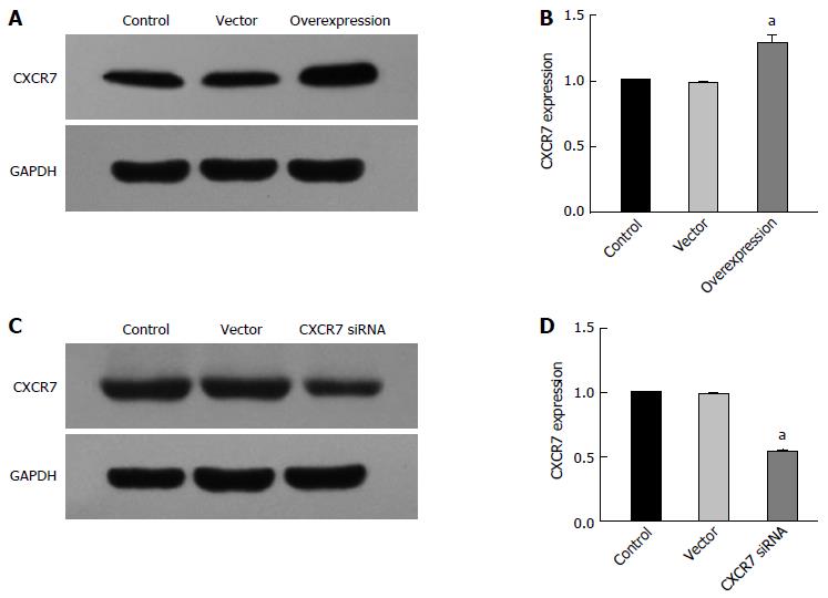
Figure 5 Stable expression of CXCR7 in human SGC7901 cells.
A: Western blot assay showed increased CXCR7 expression in stable CXCR7-overexpressing human SGC7901 cells compared with the untreated control group and blank vector transfection control group; B: PCR assay showed the same results as the Western blot assay; C: Western blot assay showed decreased endogenous CXCR7 expression in stable CXCR7-silenced human SGC 7901 cells compared with the untreated control group and blank vector transfection control group; β-actin was used as an internal control; D: PCR assay showed the same results as the Western blot assay. aP < 0.05, vs the control and vector groups. The data are presented as the mean ± SD; n = 4; bars indicate SD, P < 0.05.
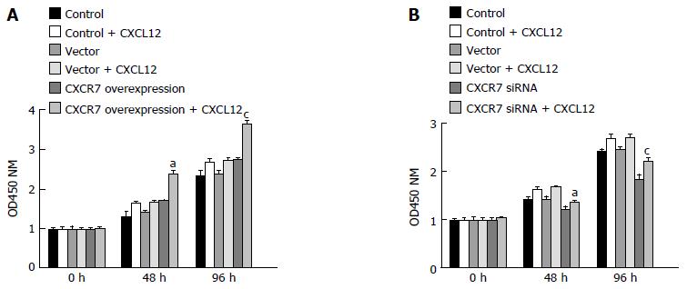
Figure 6 Effect of CXCR7 on proliferation of human gastric SGC7901 cells.
A: Overexpression of CXCR7 promoted cell proliferation; B: Silencing of CXCR7 inhibited cell proliferation. Cell proliferation was measured by CCK-8 assay at 0, 48 and 96 h. aP < 0.05, vs the control group; and cP < 0.05, vs blank vector group. Data are presented as the mean ± SD; n = 5; bars indicate SD, P < 0.05.
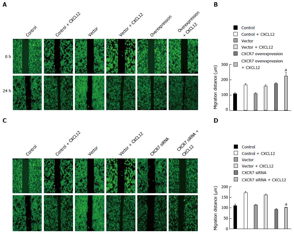
Figure 7 Effect of CXCR7 on migration of human gastric SGC7901 cells.
A: Overexpression of CXCR7 promoted CXCL12 induced enhancement of SGC-7901 cell migration in vitro; B: Mean wound width from three independent fields/wells is indicated; C: Silencing of CXCR7 inhibits CXCL12 induced enhancement of SGC-7901 cell migration in vitro; D: Mean wound width from three independent fields/wells is indicated. aP < 0.05, vs the control group and blank vector group. The data are presented as the mean ± SD; n = 3; bars indicate SD.
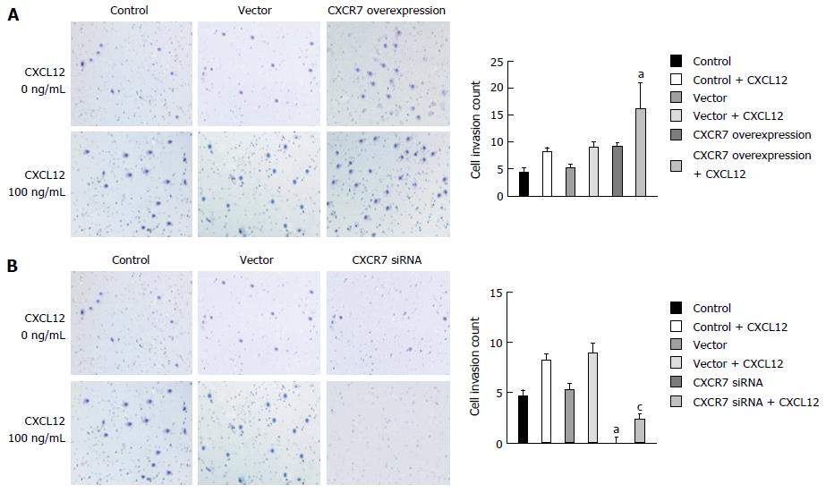
Figure 8 Effect of CXCR7 on invasion of human gastric SGC7901 cells.
A: Overexpression of CXCR7 promoted CXCL12 induced enhancement of SGC-7901 cell invasion in vitro; B: Silencing of CXCR7 decreased CXCL12 induced enhancement of SGC-7901 cell invasion in vitro. aP < 0.05, vs the control group; and cP < 0.05, vs blank vector group. The data are presented as the mean ± SD; n = 3; bars indicate SD, P < 0.05.
- Citation: Xin Q, Zhang N, Yu HB, Zhang Q, Cui YF, Zhang CS, Ma Z, Yang Y, Liu W. CXCR7/CXCL12 axis is involved in lymph node and liver metastasis of gastric carcinoma. World J Gastroenterol 2017; 23(17): 3053-3065
- URL: https://www.wjgnet.com/1007-9327/full/v23/i17/3053.htm
- DOI: https://dx.doi.org/10.3748/wjg.v23.i17.3053


