Copyright
©The Author(s) 2017.
World J Gastroenterol. Apr 14, 2017; 23(14): 2556-2565
Published online Apr 14, 2017. doi: 10.3748/wjg.v23.i14.2556
Published online Apr 14, 2017. doi: 10.3748/wjg.v23.i14.2556
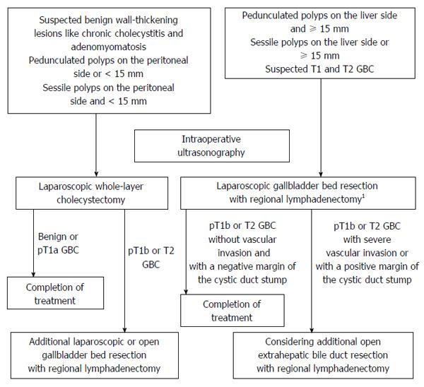
Figure 1 Algorithm of our laparoscopic approach to gallbladder lesions.
1D2 lymphadenectomy for suspected T2 GBC, and D1 lymphadenectomy for the others. D1 lymphadenectomy is defined as removal of the lymph nodes around the cystic duct and the common bile duct. D2 lymphadenectomy is defined as removal of the lymph nodes in hepatoduodenal ligament, around the common hepatic artery, and around the posterosuperior region of the pancreas head. GBC: Gallbladder carcinoma.
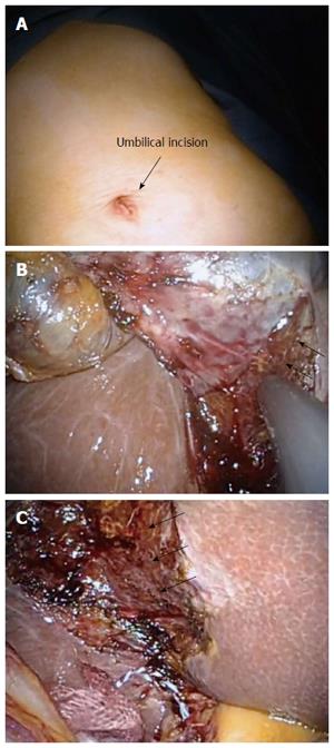
Figure 2 Surgical procedure for laparoscopic whole-layer cholecystectomy.
A: The wound just after single-incision laparoscopic whole-layer cholecystectomy; B: Detachment of the whole-layer gallbladder wall from the liver bed, leaving Laennec’s capsule (arrow) on the liver surface; C: After resection of the gallbladder.
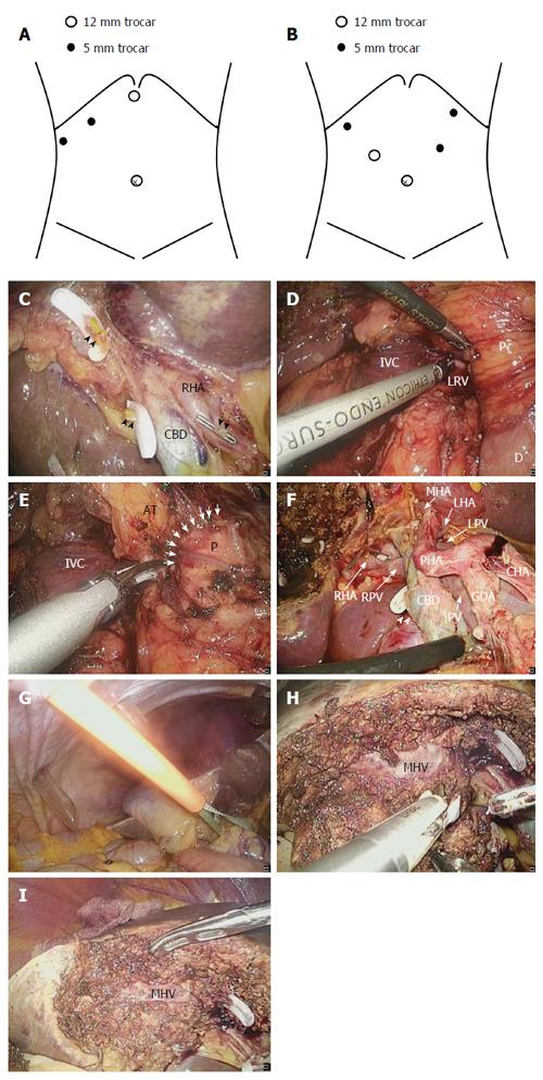
Figure 3 Surgical procedure for laparoscopic gallbladder bed resection.
A: Position of trocars in laparoscopic gallbladder bed resection (LCGB) with D1 lymphadenectomy; B: Position of trocars in LCGB with D2 lymphadenectomy; C: The cystic artery and duct are cut at their origin; D: Kocher’s mobilization; E: Lymph node dissection around the posterosuperior region of the pancreas head. Arrow indicates the boundary between the pancreatic parenchyma and surrounding adipose tissues; F: Completion of D2 lymphadenectomy; G: Performance of the Pringle maneuver with an extracorporeal tourniquet; H: Transection of the liver parenchyma by the clamp crushing method; I: After the gallbladder bed resection. RHA: Right hepatic artery; CBD: Common bile duct; Arrowhead: Stump of the cystic duct; Dotted arrow: Stump of the cystic artery; P: Pancreatic head; D: Duodenum; IVC: Inferior vena cava; LRV: Left renal vein; AT: Adipose tissues; GDA: Gastroduodenal artery; CHA: Common hepatic artery; PHA: Proper hepatic artery; LHA: Left hepatic artery; MHA: Middle hepatic artery; PV: Portal vein; LPV: Left portal vein; RPV: Right portal vein; MHV: Middle hepatic vein.
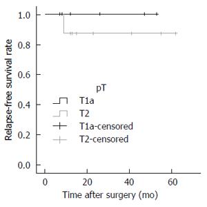
Figure 4 Relapse-free survival rate after laparoscopic surgery for pathologically diagnosed T1a and T2 gallbladder carcinoma.
- Citation: Ome Y, Hashida K, Yokota M, Nagahisa Y, Okabe M, Kawamoto K. Laparoscopic approach to suspected T1 and T2 gallbladder carcinoma. World J Gastroenterol 2017; 23(14): 2556-2565
- URL: https://www.wjgnet.com/1007-9327/full/v23/i14/2556.htm
- DOI: https://dx.doi.org/10.3748/wjg.v23.i14.2556


