Copyright
©The Author(s) 2017.
World J Gastroenterol. Apr 7, 2017; 23(13): 2337-2345
Published online Apr 7, 2017. doi: 10.3748/wjg.v23.i13.2337
Published online Apr 7, 2017. doi: 10.3748/wjg.v23.i13.2337
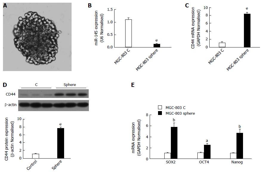
Figure 1 miR-145 and CD44 expression in gastric cancer cells with self-renewal properties.
aP < 0.05, bP < 0.01, eP < 0.001 vs the monolayer cells; data are the mean ± SEM of at least three independent experiments. A: Representative image of a tumor sphere of MGC-803 cells. MGC-803 cells were cultured in stem cell medium as described in the Materials and Methods section; B: miR-145 expression in tumor spheres and monolayer cells. miR-145 expression was determined by quantitative real-time polymerase chain reaction (qPCR); C: CD44 mRNA expression in tumor spheres and monolayer cells. CD44 mRNA expression was determined by qPCR; D: CD44 protein expression in tumor spheres and monolayer cells. Cells were harvested for western blotting analysis; E: The expression of several gastric cancer stem cell markers in tumor spheres and monolayer cells. Sox2, Oct4, and Nanog mRNA expression was determined by real-time PCR.
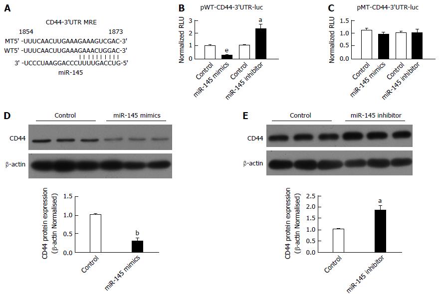
Figure 2 miR-145 targets the CD44 3’UTR directly in gastric cancer cells.
aP < 0.05, bP < 0.01, eP < 0.001 vs the control; data are the mean ± SEM of at least three independent experiments. A: A putative miRNA-recognition element (MRE) for miR-145 on the 3’UTR of CD44; B: miR-145 regulated CD44 3’UTR activity negatively; C: MRE site-mutation abolished the effects of miR-145 on CD44 3’UTR activity. MGC-803 cells were co-transfected with pMT-CD44-3’ UTR-luc or pWT-CD44-3’UTR-luc with or without miR-145 mimics or an miR-145 inhibitor, respectively, and the transfected cells were harvested 36 h later for luciferase reporter assays as described; D: miR-145 mimics inhibited CD44 protein expression; E: The miR-145 inhibitor increased CD44 protein expression. MGC-803 cells were transfected with or without miR-145 mimics or a miR-145 inhibitor, respectively, and the transfected cells were harvested 48 h later for western blotting analysis. RLU: Relative luciferase activity.
Figure 3 Overexpression of CD44 abolished the inhibitory effect of miR-145 on tumor sphere formation in gastric cancer cells.
eP < 0.001 vs cells transfected with the control; bP < 0.01 vs cells transfected with miR-145 mimics; data are the mean ± SEM of at least three independent experiments. A: Transfection with plasmid pLV-CD44 increased CD44 protein expression significantly; B: CD44 overexpression abolished the inhibitory effect of miR-145 on tumor sphere formation. MGC-803 cells were co-transfected with pLV-CD44 with or without miR-145 mimics, respectively, and the transfected cells were collected 12 h later for tumor sphere formation assays as described.
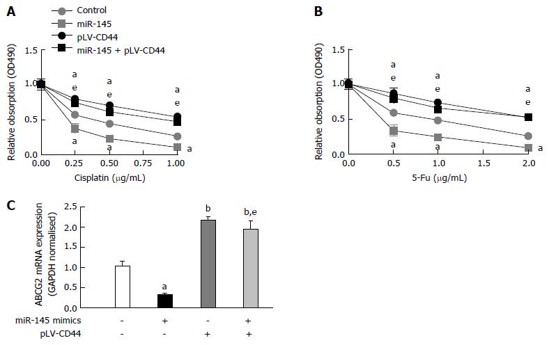
Figure 4 Overexpression of CD44 abolishes the chemo-resistance lowering effect of miR-145 in gastric cancer cells.
aP < 0.05, bP < 0.01 vs cells transfected with the control; eP < 0.001 vs cells transfected with miR-145 mimics; data are the mean ± SEM of at least three independent experiments. A: CD44 overexpression reduced MGC-803 cell chemo-resistance to cisplatin; B: CD44 overexpression reduced MGC-803 cell chemo-resistance to 5-FU. MGC-803 cells were co-transfected with pLV-CD44 with or without miR-145 mimics, respectively, the transfected cells were collected 12 h later, and then various concentrations of cisplatin or 5-FU were used to treat the cells. Cell viability was determined as described; C: ABCG2 expression following transfection of miR-145 or pLV-CD44. MGC-803 cells were co-transfected with pLV-CD44 with or without miR-145 mimics, respectively, the transfected cells were collected 36 h later, and ABCG2 mRNA expression was determined by quantitative real-time polymerase chain reaction.
- Citation: Zeng JF, Ma XQ, Wang LP, Wang W. MicroRNA-145 exerts tumor-suppressive and chemo-resistance lowering effects by targeting CD44 in gastric cancer. World J Gastroenterol 2017; 23(13): 2337-2345
- URL: https://www.wjgnet.com/1007-9327/full/v23/i13/2337.htm
- DOI: https://dx.doi.org/10.3748/wjg.v23.i13.2337


