Copyright
©The Author(s) 2017.
World J Gastroenterol. Mar 28, 2017; 23(12): 2251-2257
Published online Mar 28, 2017. doi: 10.3748/wjg.v23.i12.2251
Published online Mar 28, 2017. doi: 10.3748/wjg.v23.i12.2251
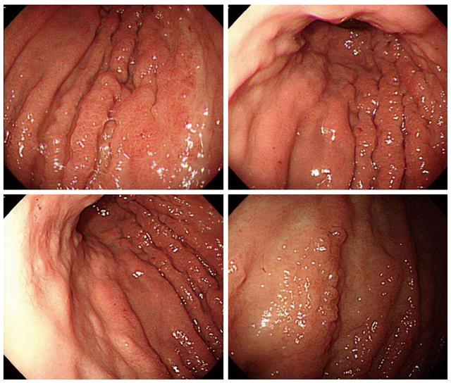
Figure 1 Upper endoscopy shows diffuse infiltrative mucosal lesion with extensive nodular thickening of the stomach wall, involving lower two-thirds of body.
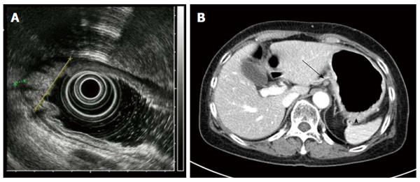
Figure 2 Endoscopic ultrasound shows subserosal invasion of the gastric lesion with lymph node involvement (A, B).
Abdomen CT scan shows infiltrative gastric lesion involving cardia and angle of stomach (arrowhead) with enlarged perigastric lymph node (arrow).
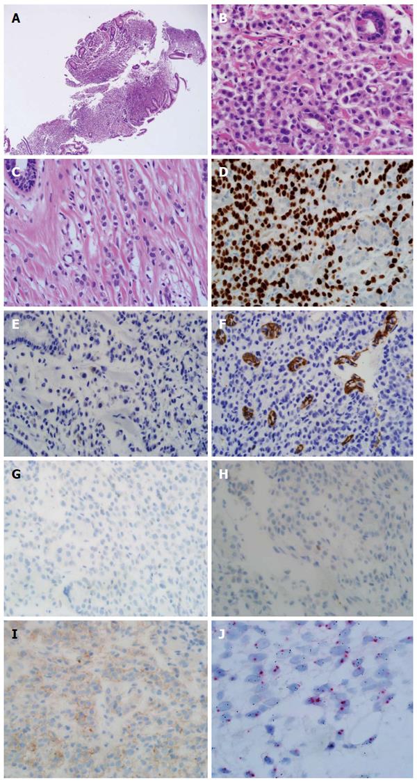
Figure 3 Pathologic features of endoscopic biopsy specimen.
Discohesive tumor cells are infiltrated in the stroma of the stomach mucosal tissue (HE × 40, A). Tumor cells show enlarged centrally located nucleus without intracytoplasmic clear mucin. The tumor cells had no connection to the remained normal gastric mucosal tissue (HE × 400, B). Previous breast cancer pathology was reviewed (C). Discohesive tumor cells were arranged in indian file. The tumor cells had enlarged centrally located nucleus without intracytoplasmic mucin (HE × 400, C). Immunohistochemical stains and molecular test of tumor was done (D-J). Diffuse strong nucleus expression of GATA3 was observed (GATA3 × 400, D). Focal, less than one percentage cytoplasmic expression of GCDFP was detected (GCDFP × 400, E). Negative stain for E-cadherin (E-cadherin × 400, F). Negative stains for ER and PR (ER × 400, PR × 400, G, H). Immunohistochemical stain for HER-2 was equivocal (HER-2 × 400, I). Silver in situ hybridization (SISH) for determination of HER2 gene status. Occasional HER2 gene amplified cells were noted in the mixture with normal HE2 gene expressing cells (SISH × 1000, J).
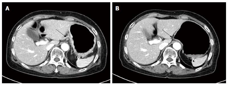
Figure 4 Response evaluation after 2 cycles of docetaxel chemotherapy (A, B).
Abdominal CT scan shows decreased perigastric lymph modes (arrows) and gastric mucosal thickening (arrowheads).
- Citation: Yim K, Ro SM, Lee J. Breast cancer metastasizing to the stomach mimicking primary gastric cancer: A case report. World J Gastroenterol 2017; 23(12): 2251-2257
- URL: https://www.wjgnet.com/1007-9327/full/v23/i12/2251.htm
- DOI: https://dx.doi.org/10.3748/wjg.v23.i12.2251


