Copyright
©The Author(s) 2017.
World J Gastroenterol. Mar 28, 2017; 23(12): 2168-2174
Published online Mar 28, 2017. doi: 10.3748/wjg.v23.i12.2168
Published online Mar 28, 2017. doi: 10.3748/wjg.v23.i12.2168
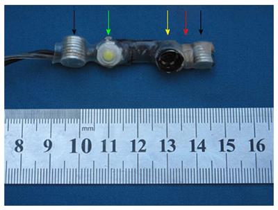
Figure 1 The miniature magnetically anchored camera with a 30° downward angle.
It consists of inner magnets (black arrow), a light source (green arrow), a vision model (yellow arrow), and a metal hexagonal nut (red arrow).
Figure 2 In vitro test.
A: The bench test consists of a special mannequin and a laparoscope; B: External view image for trocar-less laparoscopic cholecystectomy in vitro: the external magnetically anchored unit (red arrow) and the vision output device (yellow arrow); C: Critical view image for trocar-less laparoscopic cholecystectomy in vitro (the picture-in-picture view is the image captured by the miniature magnetically anchored camera with its own light source).
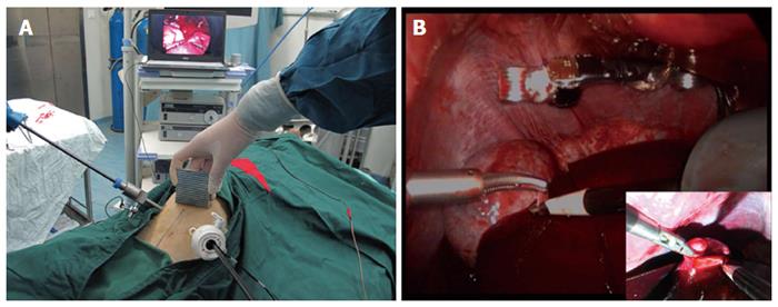
Figure 3 In vivo test.
A: External view image for trocar-less laparoscopic cholecystectomy in a canine mode; B: Critical view image for trocar-less laparoscopic cholecystectomy in a canine model (the picture-in-picture view is the image captured by the miniature magnetically anchored camera with its own light source).
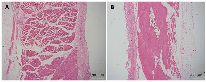
Figure 4 Pathologic assessment of abdominal wall.
A: Hematoxylin-eosin (HE) staining of normal area; B: HE staining of active area.
Figure 5 The method of achieving a 30° downward angle using the miniature magnetically anchored camera.
- Citation: Dong DH, Zhu HY, Luo Y, Zhang HK, Xiang JX, Xue F, Wu RQ, Lv Y. Miniature magnetically anchored and controlled camera system for trocar-less laparoscopy. World J Gastroenterol 2017; 23(12): 2168-2174
- URL: https://www.wjgnet.com/1007-9327/full/v23/i12/2168.htm
- DOI: https://dx.doi.org/10.3748/wjg.v23.i12.2168


