Copyright
©The Author(s) 2016.
World J Gastroenterol. Mar 7, 2016; 22(9): 2647-2656
Published online Mar 7, 2016. doi: 10.3748/wjg.v22.i9.2647
Published online Mar 7, 2016. doi: 10.3748/wjg.v22.i9.2647
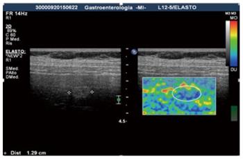
Figure 1 Image of a focal liver lesion obtained by static real-time elastography: The focal liver lesion appears hyperechoic at ultrasound image (left) and stiffer (predominantly blue) in the elasticity image.
At histology the lesion revealed to be a hepatocellular carcinoma.
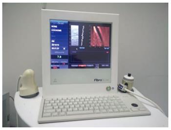
Figure 2 Transient elastography uses a low-frequency pulsed excitation to generate shear waves in the tissue, with the velocity of the shear wave - established to be related to tissue stiffness - to be measured with an ultrasound pulse-echo technique and used to calculate the elasticity.
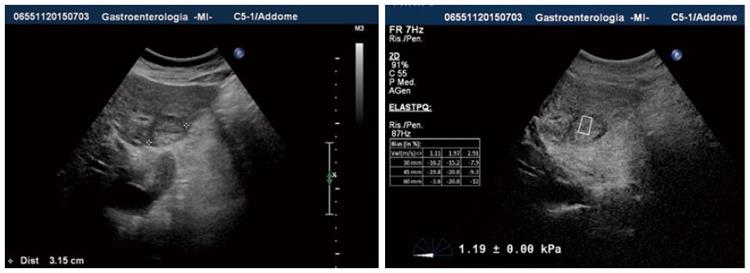
Figure 3 Shear wave ultrasound elastography allows to gather information on the tissue stiffness of both the parenchyma and focal liver lesion, obtaining a quantitative assessment.
- Citation: Conti CB, Cavalcoli F, Fraquelli M, Conte D, Massironi S. Ultrasound elastographic techniques in focal liver lesions. World J Gastroenterol 2016; 22(9): 2647-2656
- URL: https://www.wjgnet.com/1007-9327/full/v22/i9/2647.htm
- DOI: https://dx.doi.org/10.3748/wjg.v22.i9.2647


