Copyright
©The Author(s) 2016.
World J Gastroenterol. Feb 28, 2016; 22(8): 2533-2544
Published online Feb 28, 2016. doi: 10.3748/wjg.v22.i8.2533
Published online Feb 28, 2016. doi: 10.3748/wjg.v22.i8.2533
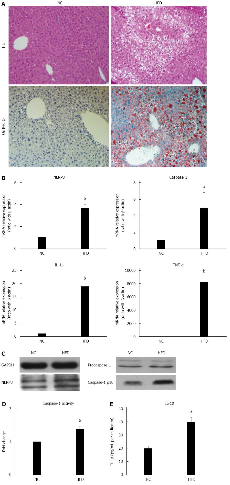
Figure 1 High fat diets induce hepatic steatosis and NOD-like receptor protein 3 inflammasome activation.
Wild-type C57BL/6 mice were fed either normal control (NC) diet or high-fat diet (HFD) for 14 wk. A: Liver histology (magnification × 100); B: mRNA expression of NLRP3 inflammasome components and proinflammatory cytokines, determined by quantitative real-time PCR; C: Protein expression of NLRP3 and caspase-1 in Western blot; D: Caspase-1 activity in liver tissue; and E: Mature IL-1β secretion in liver. Values are Mean ± SEM, aP < 0.05, bP < 0.01 vs NC; n = 10 animals per group. NLRP3: NOD-like receptor protein 3; GAPDH: Reduced glyceraldehyde-phosphate dehydrogenase; IL-1β: Interleukine-1 beta.
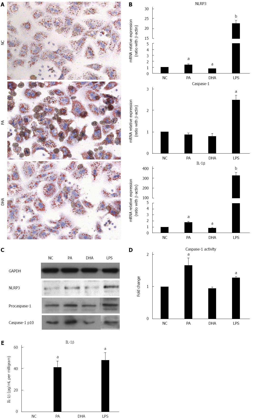
Figure 2 Effect of different fatty acids on NOD-like receptor protein 3 inflammasome activation in hepatocytes.
Primary hepatocytes were isolated from wild-type C57BL/6 mice and were treated with either palmitic acid (PA, 0.5 mmol/L) or docosahexaenoic acid (DHA, 50 μmol/L). A: Lipid deposition in hepatocytes after exposure to PA or DHA for 24 h; B: mRNA expression of NLRP3 inflammasome components after FFAs treatment for 6 h; C: Protein expression of NLRP3 and caspase-1 in Western blot; D: Caspase-1 activity and E: Culture supernatant IL-1β level. Values are Mean ± SEM, aP < 0.05, bP < 0.01 vs normal control (NC), n = 3 experiments. NLRP3: NOD-like receptor protein 3; GAPDH: Reduced glyceraldehyde-phosphate dehydrogenase; LPS: Lipopolysaccharide; IL-1β: Interleukine-1 beta.
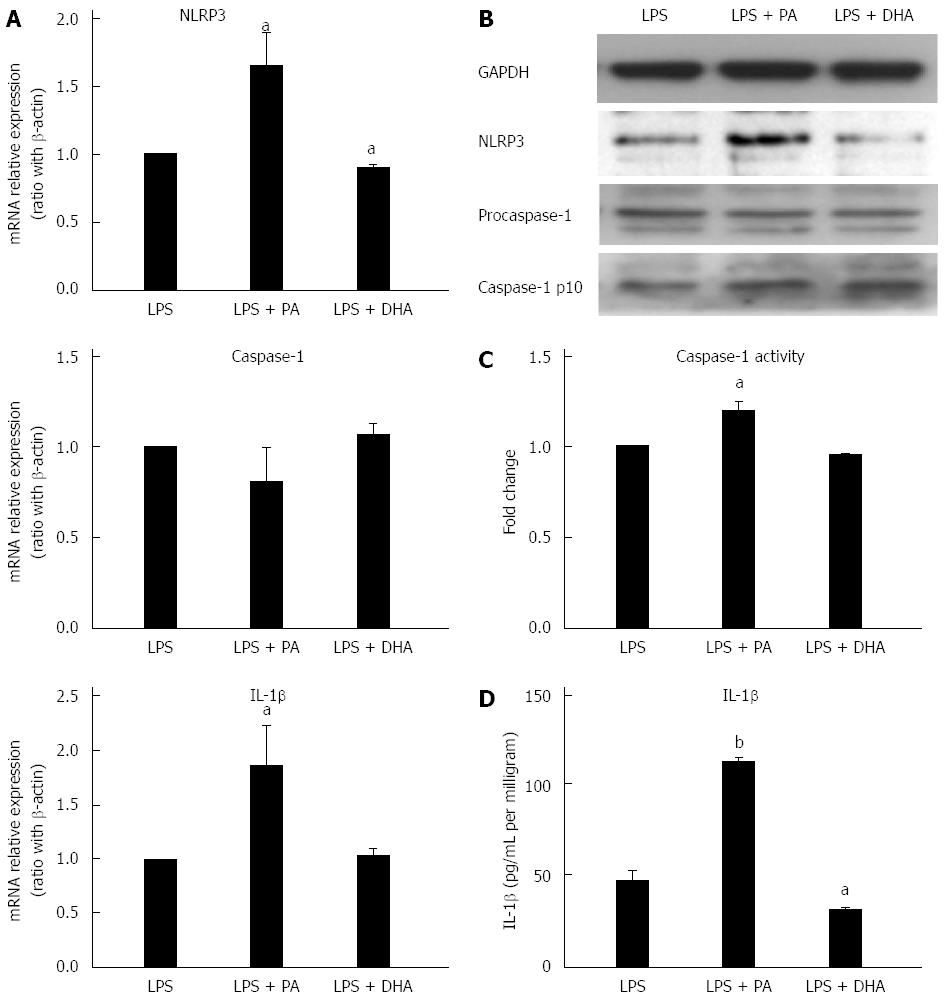
Figure 3 Combined effect of free fatty acids with lipopolysaccharide on NOD-like receptor protein 3 inflammasome activation in hepatocytes.
Primary hepatocytes were isolated and treated with either PA (0.5 mmol/L) or DHA (50 μmol/L) combined with LPS (1 μg/mL). A: mRNA expression of NOD-like receptor protein 3 (NLRP3) inflammasome components; B: Protein expression of NLRP3 and caspase-1; C: Caspase-1 activity; and D: Culture supernatant IL-1β level. Values are mean ± SEM, aP < 0.05, bP < 0.01 vs LPS control, n = 3 experiments. NF-κB: Nuclear factor-kappa B; PA: Palmitic acid; DHA: Docosahexaenoic acid; LPS: Lipopolysaccharide; FFAs: Free fatty acids; GAPDH: Reduced glyceraldehyde-phosphate dehydrogenase. IL-1β: Interleukine-1 beta
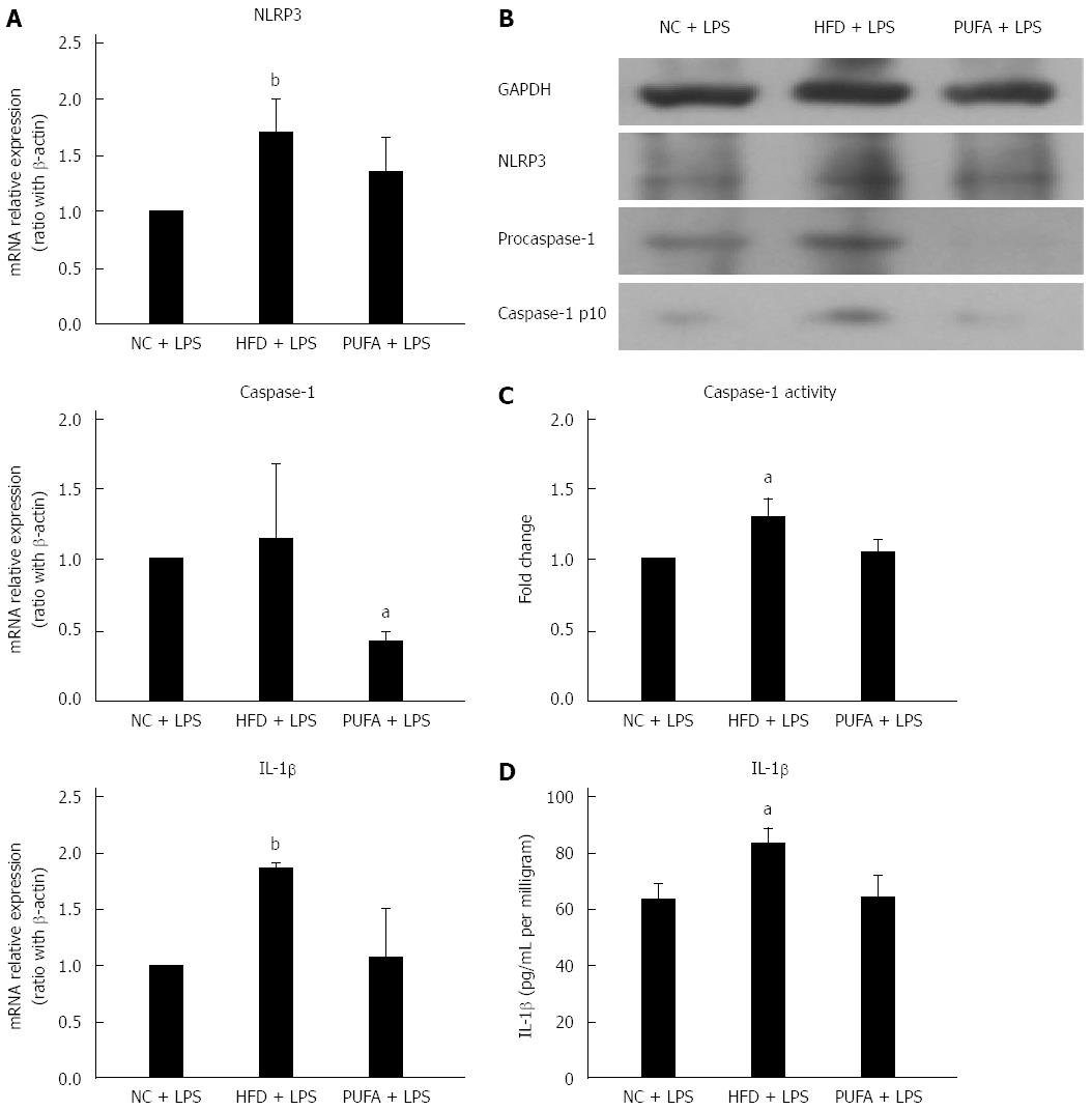
Figure 4 Effect of different fatty acid diets combined with lipopolysaccharide on hepatic NOD-like receptor protein 3 inflammasome in vivo.
Wild-type C57BL/6 mice were fed either normal control (NC) diet, high-fat diet (HFD) or PUFA-enriched diet (PUFA) for 4 wk and then injected with LPS (1.5 mg/kg) before being sacrificed. Liver tissues were obtained for the following analysis. A: mRNA expression of NLRP3 inflammasome components; B: Protein expression of NLRP3 and caspase-1; C: Caspase-1 activity; and D: Mature IL-1β secretion in liver. Values are mean ± SEM, aP < 0.05, bP < 0.01 vs NC + LPS, n = 5 animals per group. NLRP3: NOD-like receptor protein 3; GAPDH: Reduced glyceraldehyde-phosphate dehydrogenase; LPS: Lipopolysaccharide; IL-1β: Interleukine-1 beta.
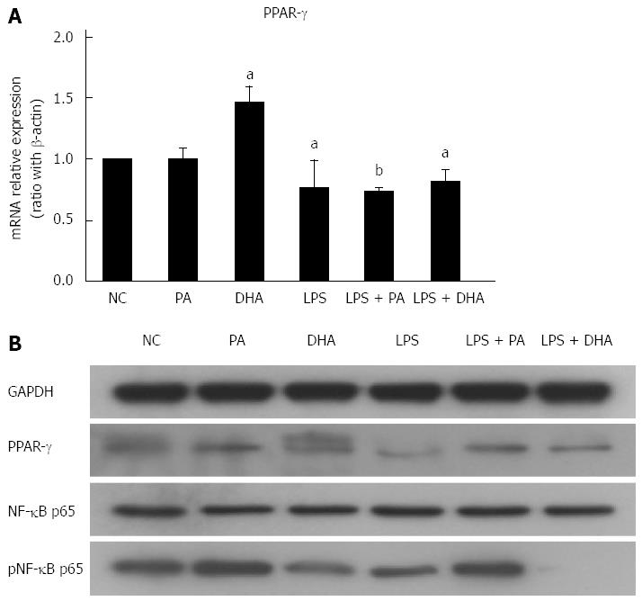
Figure 5 Effect of different fatty acids on nuclear factor-kappa B/peroxisome proliferator-activated receptor-γ expression.
Primary hepatocytes were isolated and treated with PA (0.5 mmol/L), DHA (50 μmol/L) or LPS (1 μg/mL) alone or both of them. (A) mRNA expression of PPAR-γ, (B) Protein expression of NF-κB and PPAR-γ. Values are mean ± SEM, aP < 0.05, bP < 0.01 vs control, n = 3 experiments. PPAR-γ: Peroxisome proliferator-activated receptor-γ; NF-κB: Nuclear factor-kappa B; PA: Palmitic acid; DHA: Docosahexaenoic acid; LPS: Lipopolysaccharide.
- Citation: Sui YH, Luo WJ, Xu QY, Hua J. Dietary saturated fatty acid and polyunsaturated fatty acid oppositely affect hepatic NOD-like receptor protein 3 inflammasome through regulating nuclear factor-kappa B activation. World J Gastroenterol 2016; 22(8): 2533-2544
- URL: https://www.wjgnet.com/1007-9327/full/v22/i8/2533.htm
- DOI: https://dx.doi.org/10.3748/wjg.v22.i8.2533


