Copyright
©The Author(s) 2016.
World J Gastroenterol. Feb 21, 2016; 22(7): 2383-2390
Published online Feb 21, 2016. doi: 10.3748/wjg.v22.i7.2383
Published online Feb 21, 2016. doi: 10.3748/wjg.v22.i7.2383
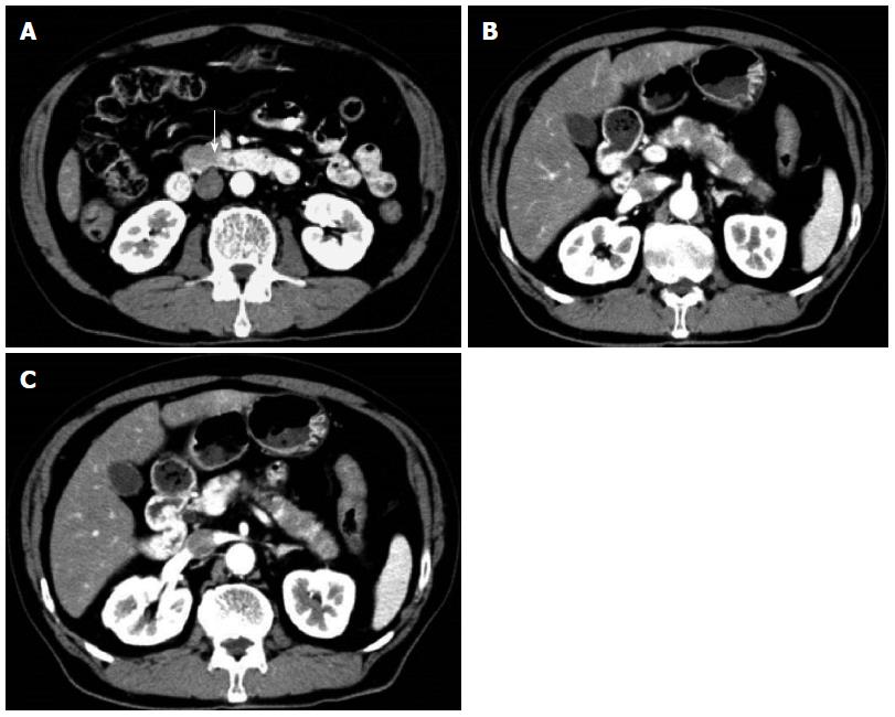
Figure 1 Contrast-enhanced abdominal/pelvic computed tomography showing pancreatic enlargement with a diffuse, poorly enhanced area in the uncinate process (arrow) and pancreatic body tail (A-C).
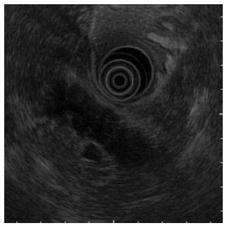
Figure 2 On endoscopic ultrasound, a hypoechoic mass in a form in which the pancreatic lobe structure was maintained in the uncinate process and from the pancreatic body to the tail.
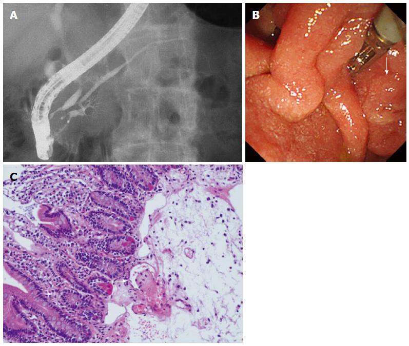
Figure 3 Endoscopic retrograde pancreatography.
A: A slight disparity of the opening diameter in the main pancreatic duct, but no localized stenosis or diffuse narrowing observed; B: No abnormality in the papilla of Vater (arrow); C: The biopsy reveals thrombus formation (arrow).
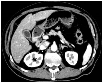
Figure 4 Contrast-enhanced abdominal/pelvic computed tomography performed in May 2014 showing a trend of improvement in the pancreatic enlargement.
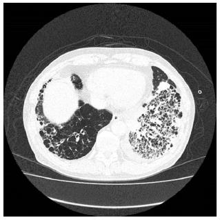
Figure 5 Chest computed tomography demonstrating an alveolar hemorrhage and pneumonia.
- Citation: Iida T, Adachi T, Tabeya T, Nakagaki S, Yabana T, Goto A, Kondo Y, Kasai K. Rare type of pancreatitis as the first presentation of anti-neutrophil cytoplasmic antibody-related vasculitis. World J Gastroenterol 2016; 22(7): 2383-2390
- URL: https://www.wjgnet.com/1007-9327/full/v22/i7/2383.htm
- DOI: https://dx.doi.org/10.3748/wjg.v22.i7.2383


