Copyright
©The Author(s) 2016.
World J Gastroenterol. Dec 28, 2016; 22(48): 10566-10574
Published online Dec 28, 2016. doi: 10.3748/wjg.v22.i48.10566
Published online Dec 28, 2016. doi: 10.3748/wjg.v22.i48.10566
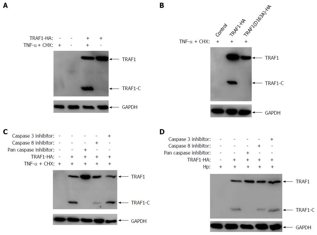
Figure 1 Tumor necrosis factor receptor-associated factor 1 is cleaved via caspase-8 in AGS cells infected with Helicobacter pylori.
A: AGS cells transfected with either HA-tagged tumor necrosis factor receptor-associated factor 1 (TRAF1) or empty vector were treated with the apoptosis inducer (0.3 μg/mL CHX and 80 ng/mL TNF-α) for 6 h, and the cleavage of TRAF1 was detected by western blotting; B: AGS cells were transfected with wild-type or D163A mutant of TRAF1 and analyzed for the cleavage of TRAF1 by western blotting; C: AGS cells transfected with the HA-tagged TRAF1 were pretreated with 20 μmol/L of Z-VAD-FMK, Z-IETD-FMK, or Z-YVAD-FMK for 30 min, followed by treatment with the apoptosis inducer for 6 h and then analyzed for the cleavage of TRAF1 by western blotting; D: AGS cells transfected with the HA-tagged TRAF1 were pretreated with 20 μmol/L of Z-VAD-FMK, Z-IETD-FMK, or Z-YVAD-FMK for 30 min, followed by infection with Helicobacter pylori strain NCTC11637 for 6 h and then analyzed for the cleavage of TRAF1 by western blotting.
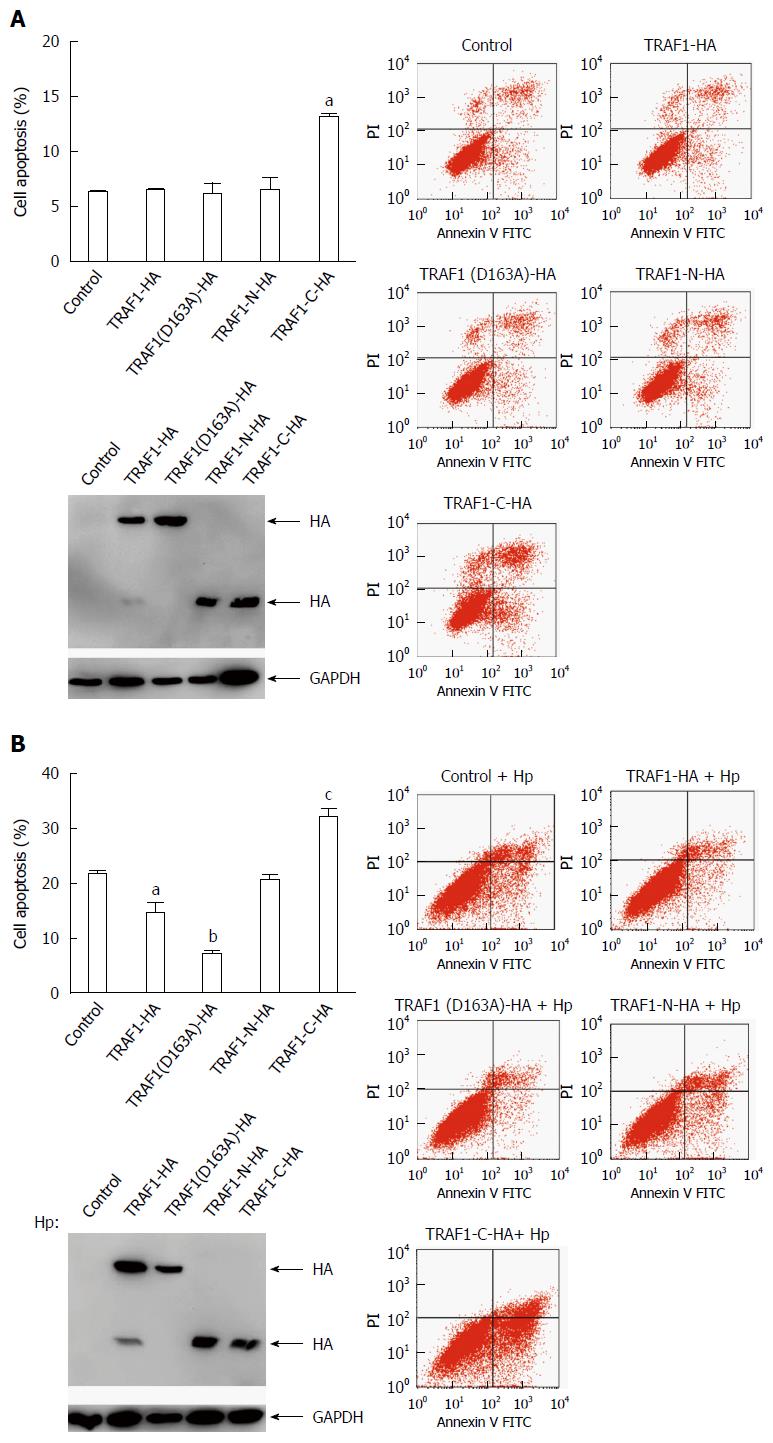
Figure 2 COOH-terminal tumor necrosis factor receptor-associated factor 1 fragment promoted cell apoptosis in Helicobacter pylori infected cells.
A: AGS cells were transfected with HA-tagged tumor necrosis factor receptor-associated factor 1 (TRAF1), TRAF1(D163A), TRAF1-N, TRAF1-C for 24 h and then analyzed for cell apoptosis by flow cytometry using annexin V-FITC and propidium iodide staining (aP < 0.05 vs control). The expression levels of these proteins in the analyzed cells were shown (Lower); B: AGS cells were transfected with HA-tagged TRAF1, TRAF1(D163A), TRAF1-N, TRAF1-C for 24 h respectively, followed by infection with Helicobacter pylori strain NCTC11637 for 24 h and then analyzed for cell apoptosis (aP < 0.05 vs control, bP < 0.01 vs Control, cP < 0.05 vs control). The expression levels of these proteins in the analyzed cells are shown (Lower).
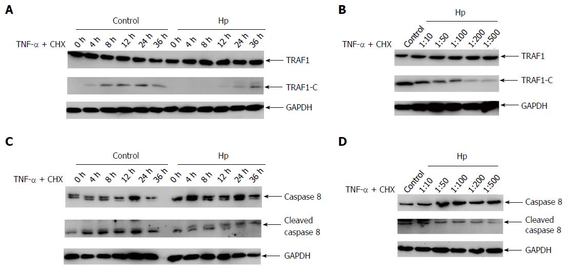
Figure 3 Helicobacter pylori inhibits the cleavage of tumor necrosis factor receptor-associated factor 1.
A: AGS cells transfected with HA-tagged tumor necrosis factor receptor-associated factor 1 (TRAF1) were treated with the apoptosis inducer (0.3 μg/mL CHX and 80 ng/mL TNF-α) and co-cultured with Helicobacter pylori (H. pylori) strain NCTC11637, or left untreated for the infection time indicated, and analyzed for the cleavage of TRAF1 by western blotting; B: AGS cells transfected with HA-tagged TRAF1 were treated with the apoptosis inducer and co-cultured with H. pylori strain NCTC11637, or left untreated, at the indicated cell/bacteria ratios and analyzed for the cleavage of TRAF1 by western blotting; C: The same as shown in (A), but analyzed for the uncleaved and cleaved caspase-8 by western blotting; D: The same as shown in (B), but analyzed for the uncleaved and cleaved caspase-8 by western blotting.
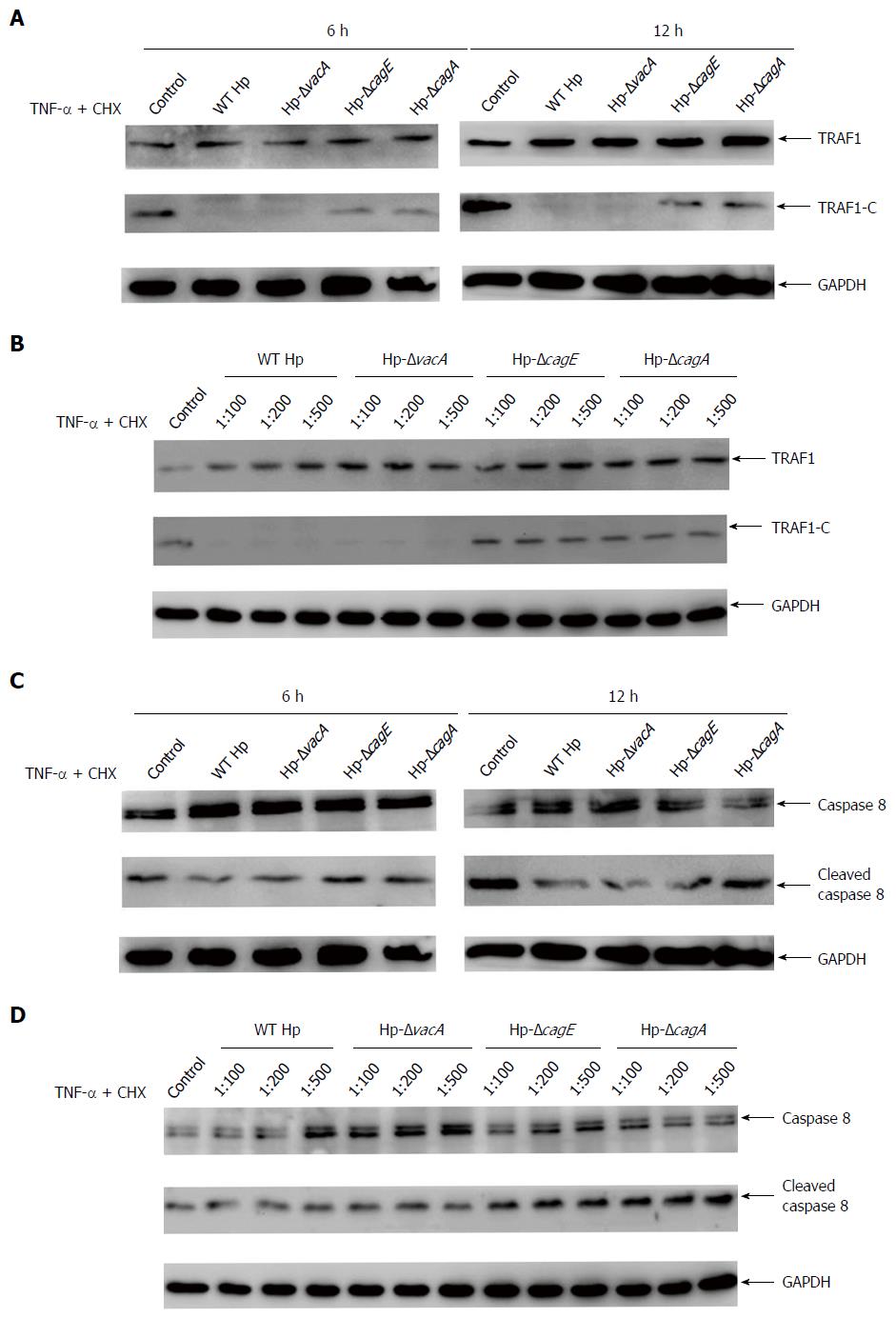
Figure 4 CagA is the major factor involved in inhibiting the cleavage of tumor necrosis factor receptor-associated factor 1.
A: AGS cells transfected with tumor necrosis factor receptor-associated factor 1 (TRAF1) were treated with the apoptosis inducer (0.3 μg/mL CHX and 80 ng/mL TNF-α), and co-cultured with Helicobacter pylori (H. pylori) strain NCTC11637 or its isogenic vacA-, cagE- or cagA-null mutants, and analyzed for the cleavage of TRAF1 at 6 h and 12 h by western blotting; B: AGS cells transfected with TRAF1 were treated with the apoptosis inducer, and co-cultured with H. pylori strain NCTC11637, or its isogenic vacA-, cagE- or cagA-null mutants at the indicated cell/bacteria ratios, and analyzed for the cleavage of TRAF1 by western blotting; C: The same as shown in (A), but analyzed for uncleaved and cleaved caspase-8 by western blotting; D: The same as shown in (B), but analyzed for uncleaved and cleaved caspase-8 by western blotting.
- Citation: Wan XK, Yuan SL, Wang YC, Tao HX, Jiang W, Guan ZY, Cao C, Liu CJ. Helicobacter pylori inhibits the cleavage of TRAF1 via a CagA-dependent mechanism. World J Gastroenterol 2016; 22(48): 10566-10574
- URL: https://www.wjgnet.com/1007-9327/full/v22/i48/10566.htm
- DOI: https://dx.doi.org/10.3748/wjg.v22.i48.10566


