Copyright
©The Author(s) 2016.
World J Gastroenterol. Dec 14, 2016; 22(46): 10260-10266
Published online Dec 14, 2016. doi: 10.3748/wjg.v22.i46.10260
Published online Dec 14, 2016. doi: 10.3748/wjg.v22.i46.10260
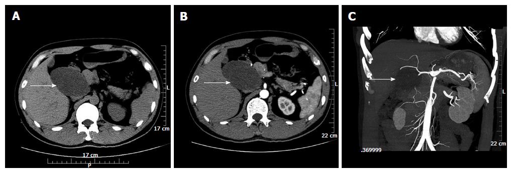
Figure 1 Computed tomography findings.
A: An unenhanced computed tomography (CT) scan showed an 8.2 cm × 5.1 cm well-defined cystic and solid mass (arrow) above the pancreatic head and adjacent to the common hepatic artery; B: On contrast-enhanced CT, the mass (arrow) showed no obvious enhancement; C: CT angiography showed that the tumor blood supply (arrow) was probably from branches of the pancreaticoduodenal artery.
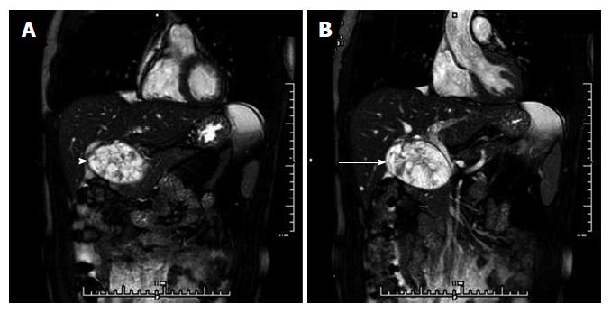
Figure 2 Magnetic resonance cholangiopancreatography findings.
A: Magnetic resonance cholangiopancreatography (MRCP) showed that the mass (arrow) was inhomogeneous and hyperintense on T2-weighted images and probably located in the pancreatic head; B: The middle-low segment of the common bile duct was compressed.
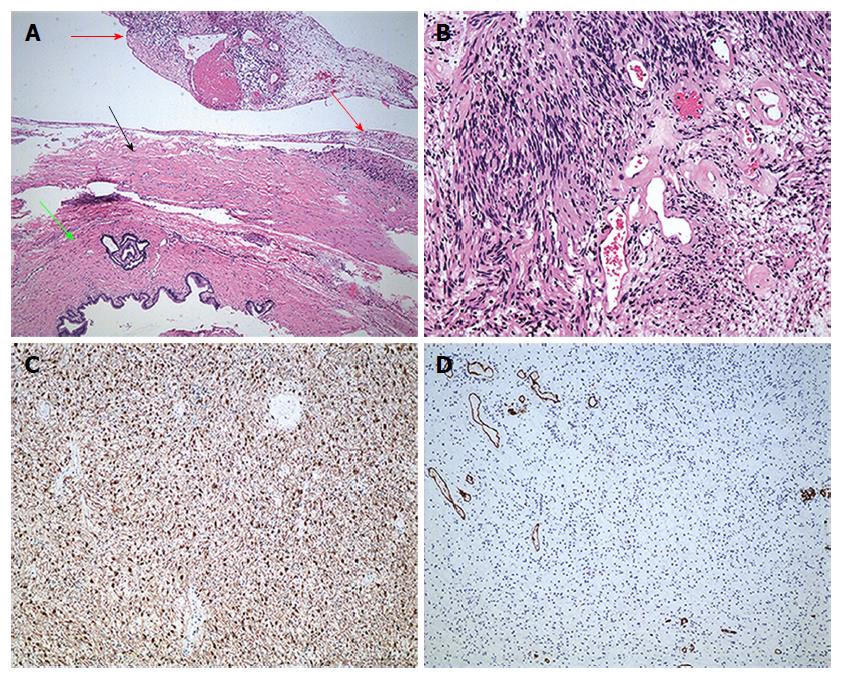
Figure 3 Microscopic examination and immunohistochemical staining.
A: Microscopically, the tumor (red arrow) with a capsule (black arrow) was adjacent to the cholecystic duct (green arrow) (HE, × 200); B: The tumor mainly consisted of spindle-shaped cells with both hypercellular and hypocellular areas (HE, × 200). Immunohistochemical investigation showed that the tumor was positive for protein S-100 (C) and negative for CD34 (D) (HE, × 100). HE: Hematoxylin and eosin.
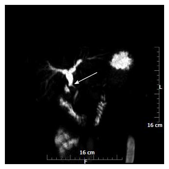
Figure 4 Magnetic resonance cholangiopancreatography findings after surgery.
Magnetic resonance cholangiopancreatography showed that the middle common bile duct segment was narrow and even interrupted (arrow), while the higher common bile duct segment and intrahepatic bile ducts were expanded.
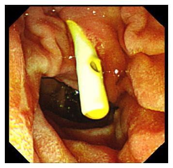
Figure 5 Endoscopic retrograde cholangiopancreatography.
Under Endoscopic retrograde cholangiopancreatography, a stent was implanted into the strictured common bile duct.
- Citation: Xu SY, Sun K, Xie HY, Zhou L, Zheng SS, Wang WL. Schwannoma in the hepatoduodenal ligament: A case report and literature review. World J Gastroenterol 2016; 22(46): 10260-10266
- URL: https://www.wjgnet.com/1007-9327/full/v22/i46/10260.htm
- DOI: https://dx.doi.org/10.3748/wjg.v22.i46.10260


