Copyright
©The Author(s) 2016.
World J Gastroenterol. Dec 14, 2016; 22(46): 10249-10253
Published online Dec 14, 2016. doi: 10.3748/wjg.v22.i46.10249
Published online Dec 14, 2016. doi: 10.3748/wjg.v22.i46.10249
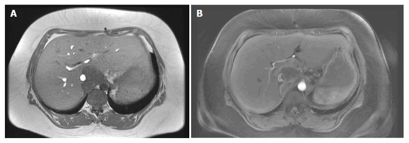
Figure 1 Magnetic resonance imaging shows an isolated liver metastasis in caudate lobe of the liver.
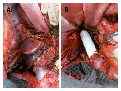
Figure 2 Distal inferior vena cava.
A: Tumor mass infiltrating the inferior vena cava (ICV) at the bifurcation of the renal veins (vessel loop); B: Vascular interposition graft after partial resection of the IVC.
- Citation: Sánchez-Velázquez P, Moosmann N, Töpel I, Piso P. “En bloc” caudate lobe and inferior vena cava resection following cytoreductive surgery and hyperthermic intraperitoneal chemotherapy for peritoneal and liver metastasis of colorectal cancer. World J Gastroenterol 2016; 22(46): 10249-10253
- URL: https://www.wjgnet.com/1007-9327/full/v22/i46/10249.htm
- DOI: https://dx.doi.org/10.3748/wjg.v22.i46.10249


