Copyright
©The Author(s) 2016.
World J Gastroenterol. Dec 7, 2016; 22(45): 10053-10063
Published online Dec 7, 2016. doi: 10.3748/wjg.v22.i45.10053
Published online Dec 7, 2016. doi: 10.3748/wjg.v22.i45.10053
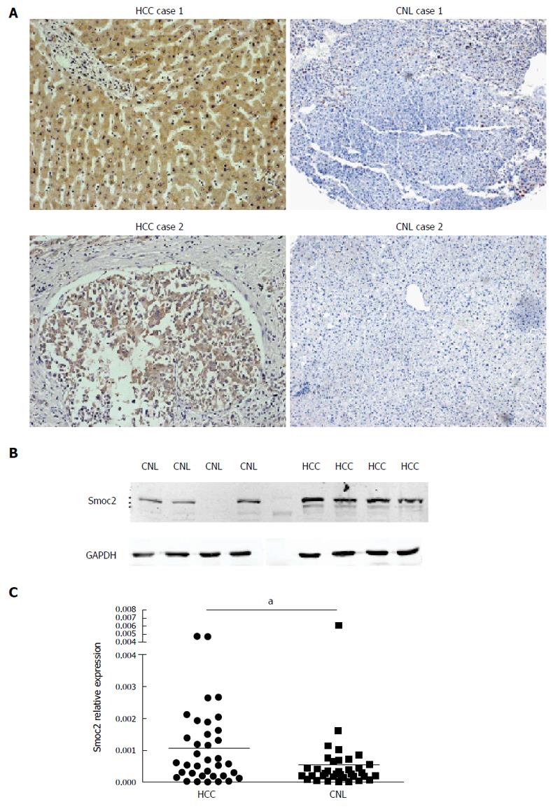
Figure 1 Smoc2 was up-regulated in hepatocellular carcinoma tissues compared with corresponding non-tumor liver tissues.
A: Representative images of immunohistochemistry (IHC) staining assay; IHC images show that expression of Smoc2 was higher in hepatocellular carcinoma (HCC) tissues compared with corresponding corresponding non-tumor liver (CNL) tissues; B: Western blot assay show the expression of Smoc2 was higher in fresh HCC tissues than in CNL tissues; C: Quantitative real-time PCR assay showed that the relative expression of Smoc2 was higher in fresh HCC tissues than in CNL tissues. aP < 0.05.
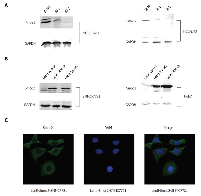
Figure 2 Western blot assay.
A: Western blot assay showing that expression of Smoc2 in MHCC-97H cells and HCC-LM3 cells was inhibited by small interfering (si)RNA; B: Western blot assay showing that expression of Smoc2 in SMMC-7721 cells and Huh7 cells was significantly up-regulated by lentivirus transfection. Lenti-Smoc2 ha overexpression of Smoc2 by lentivirus transfection; lenti-vector was the negative control of lentivirus transfection; C: Immunofluorescence staining showing overexpression of Smoc2 in SMMC-7721 cells; DAPI was used for nucleus staining.
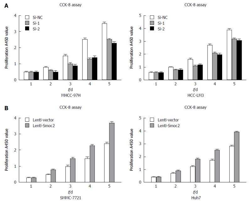
Figure 3 CCK-8 assay.
A: CCK-8 assay was used for cell viability detection and reflected cell proliferation. The results show that the proliferation ability was inhibited by small interfering (si)RNA in MHCC-97H cells and HCC-LM3 cells; B: CCK-8 assay results show that the cell proliferation ability in SMMC-7721 cells and Huh7 cells was enhanced by lenti-Smoc2-virus transfection.
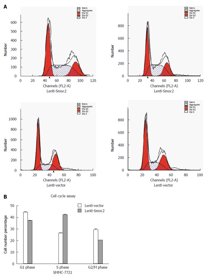
Figure 4 SMMC-7721 cells.
A: Fluorescence-activated cell sorting assay for cell cycle detection. All the SMMC-7721 cells were stained by propidium iodide, which can be detected at 488 nm; B: The overexpression of Smoc2 caused increase of S stage cell proportion in SMMC-7721 cells, which indicated promotion of cell cycle progression.
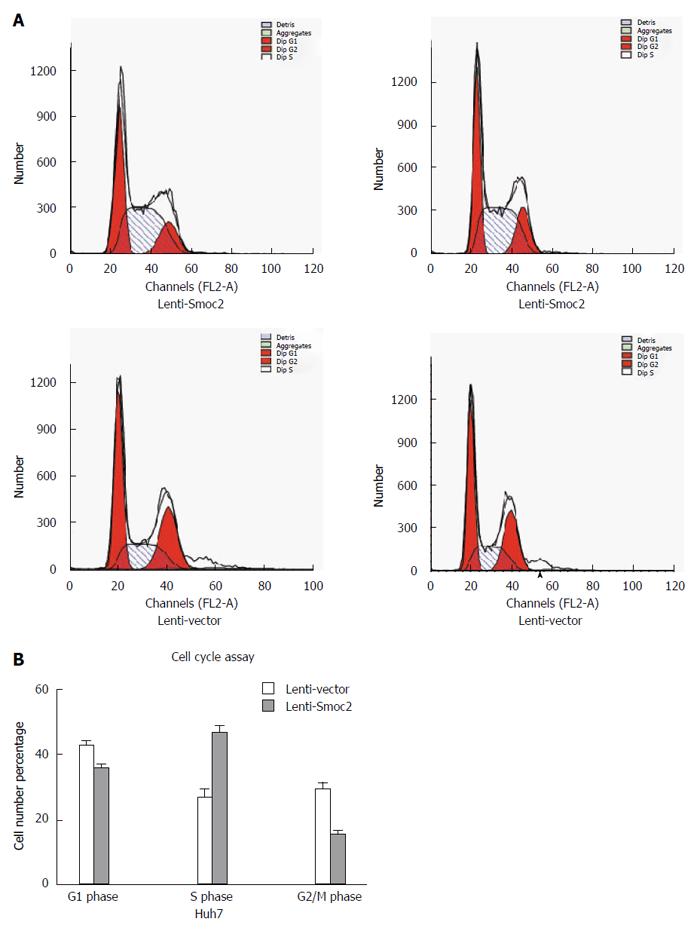
Figure 5 Huh7 cells.
A: Fluorescence-activated cell sorting assay for cell cycle detection. All the Huh7 cells were stained by propidium iodide, which can be detected at 488 nm; B: The overexpression of Smoc2 caused increase of S stage cell proportion in Huh7 cells, which indicated the promotion of cell cycle progression.
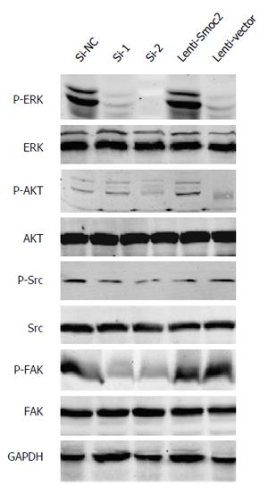
Figure 6 Phosphorylation of ERK up-regulated in Smoc2 overexpressing cells and down-regulated in cells with Smoc2 small interfering (si)RNA.
Western blot assay showed the change of phospho-ERK, Src, FAK and AKT expression by Smoc2 interference or overexpression.
- Citation: Su JR, Kuai JH, Li YQ. Smoc2 potentiates proliferation of hepatocellular carcinoma cells via promotion of cell cycle progression. World J Gastroenterol 2016; 22(45): 10053-10063
- URL: https://www.wjgnet.com/1007-9327/full/v22/i45/10053.htm
- DOI: https://dx.doi.org/10.3748/wjg.v22.i45.10053


