Copyright
©The Author(s) 2016.
World J Gastroenterol. Nov 21, 2016; 22(43): 9650-9653
Published online Nov 21, 2016. doi: 10.3748/wjg.v22.i43.9650
Published online Nov 21, 2016. doi: 10.3748/wjg.v22.i43.9650
Figure 1 Lung metastasis lesion.
A: 12 mm enhanced lung nodule was detected by initial chest computed tomography (CT); B: CT 3 mo after capecitabine; C: Initial 18F-fluorodeoxyglucose-positron emission tomography/CT.
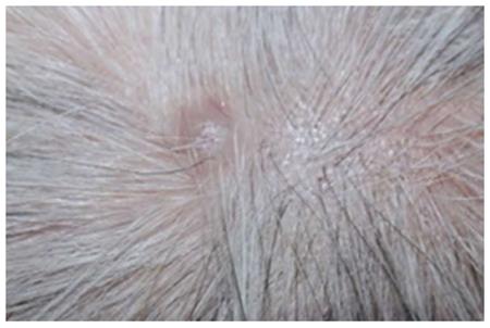
Figure 2 Cutaneous metastasis lesions in the head.
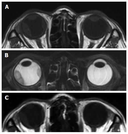
Figure 3 An elevated choroidal neoplasm.
A: T1-wieghted with low signal on the enhanced T1-weighted and higher signal on the T2-weighted image, contrast-enhanced orbital magnetic resonance imaging (MRI) with fat suppression revealed an infrabulbar mass of 12 mm × 5 mm; B: MRI T2-weighted image; C: MRI 3 mo after initiation of therapy (T1-weighed).
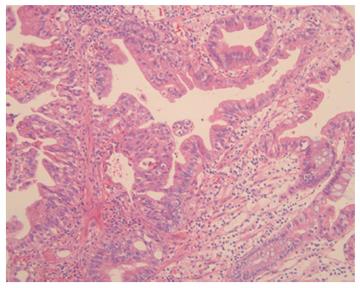
Figure 4 Pathologic findings of skin (hematoxylin-eosin staining, magnification × 100).
- Citation: Ha JY, Oh EH, Jung MK, Park SE, Kim JT, Hwang IG. Choroidal and skin metastases from colorectal cancer. World J Gastroenterol 2016; 22(43): 9650-9653
- URL: https://www.wjgnet.com/1007-9327/full/v22/i43/9650.htm
- DOI: https://dx.doi.org/10.3748/wjg.v22.i43.9650


