Copyright
©The Author(s) 2016.
World J Gastroenterol. Oct 28, 2016; 22(40): 8862-8868
Published online Oct 28, 2016. doi: 10.3748/wjg.v22.i40.8862
Published online Oct 28, 2016. doi: 10.3748/wjg.v22.i40.8862
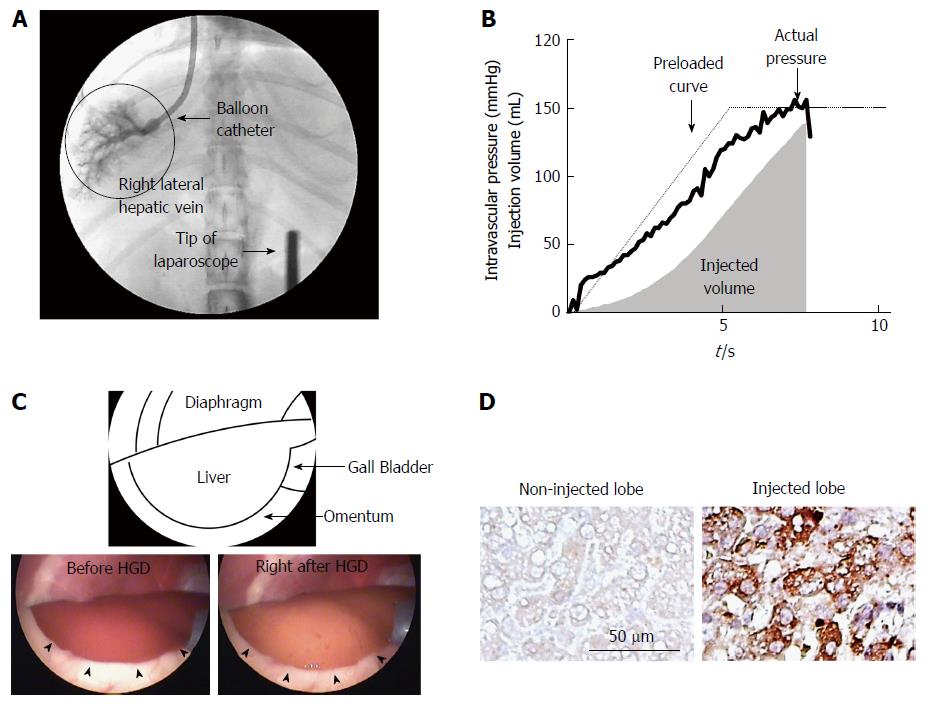
Figure 1 Image-guided, computer-controlled hydrodynamic gene-delivery to the dog liver.
The balloon catheter was placed at the appropriate position in the hepatic veins of right lateral lobe and the occlusion of the blood flow by the balloon was confirmed by injecting a small amount of contrast medium into the hepatic vein. Then the hydrodynamic injection of naked DNA solution was performed under the real time monitoring of liver structure by the laparoscope using the computer-controlled injection system (A). B: Time-pressure curve and the volume of injected solution recorded in the injection system. Solid and dotted lines represent actual and preloaded time-pressure curves. A gray area shows cumulative volume of injected saline (mL). C: Laparoscopic findings of the hydrodynamically injected right lateral lobe of the dog. The injected lobe was swollen and the injected DNA solution transiently made the liver pale. No destruction nor bleeding were seen on the surface of the liver (arrowheads). D: The effect of lobe-specific hydrodynamic gene delivery of luciferase expressing plasmid. The immunohistochemical analyses showed positively stained cells in the injected right lateral lobe. No stained cells were found in non-injected left lateral lobe.
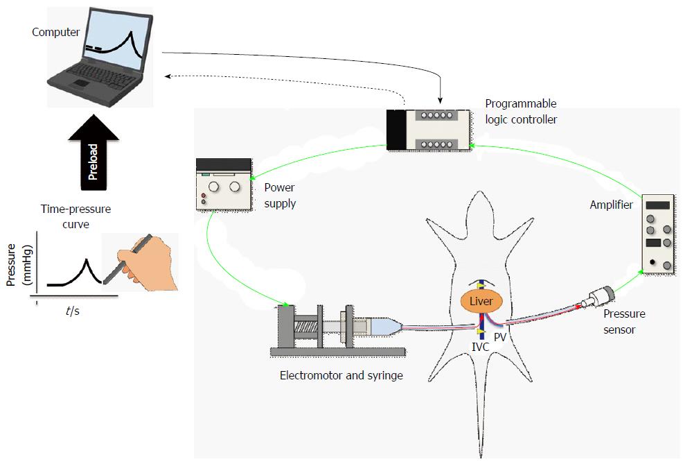
Figure 2 Scheme of computer-controlled injection system.
The schema of the newly developed hydrodynamic gene-delivery system. This figure is partly reused and modified with updated information from Figure 1 in Ref [20] with their permission. IVC: Inferior vena cava; PV: Portal vein.
- Citation: Yokoo T, Kamimura K, Abe H, Kobayashi Y, Kanefuji T, Ogawa K, Goto R, Oda M, Suda T, Terai S. Liver-targeted hydrodynamic gene therapy: Recent advances in the technique. World J Gastroenterol 2016; 22(40): 8862-8868
- URL: https://www.wjgnet.com/1007-9327/full/v22/i40/8862.htm
- DOI: https://dx.doi.org/10.3748/wjg.v22.i40.8862


