Copyright
©The Author(s) 2016.
World J Gastroenterol. Sep 28, 2016; 22(36): 8178-8186
Published online Sep 28, 2016. doi: 10.3748/wjg.v22.i36.8178
Published online Sep 28, 2016. doi: 10.3748/wjg.v22.i36.8178
Figure 1 Flow diagram of the preparation of isolated human hepatocytes with a modified four-step retrograde perfusion technique.
Buffers: PBE: Perfusion buffer with EDTA; PB: Perfusion buffer; PBD: Perfusion buffer with dispase; PBC: Perfusion buffer with collagenase; WB: Washing buffer; WBD: Washing buffer with DNase.
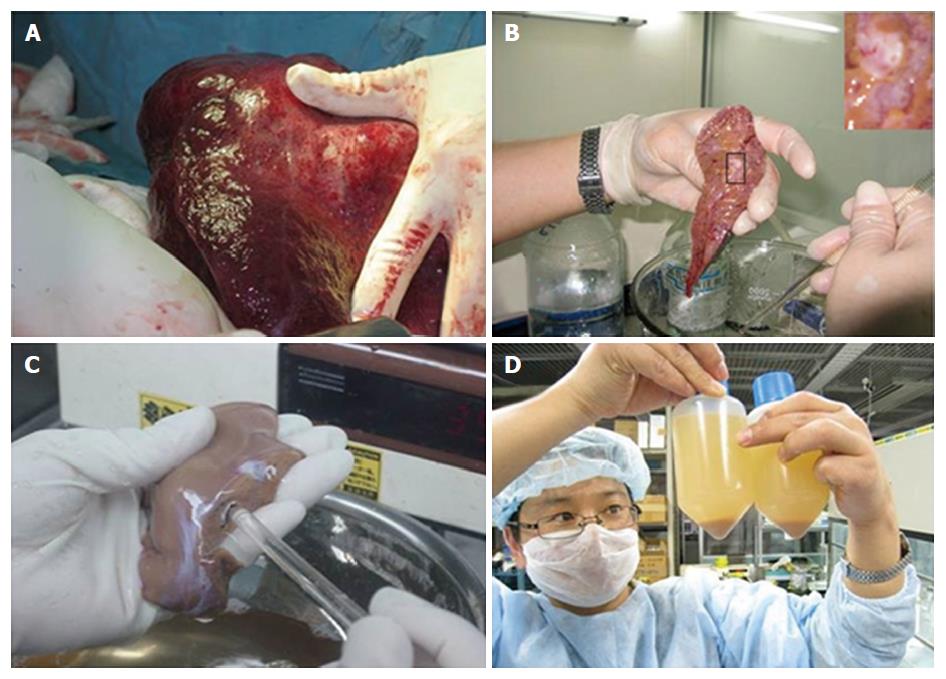
Figure 2 Preparation of liver wedges.
Only patients who had undergone left hemi-hepatectomies were deemed suitable for obtaining normal resected liver tissue from (A). Cut-off end of a suitable pipet tip to obtain an optimal size to match the vessel opening and cannulate the chosen vessel (B, C). Primary human hepatocytes must be isolated under stringent and rigorous sterile conditions (D).
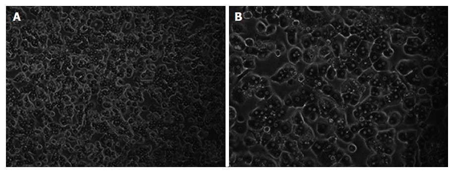
Figure 3 Phase-contrast photographs of primary human hepatocytes at 24 h after isolation.
Magnification × 100 (A) and × 200 (B).
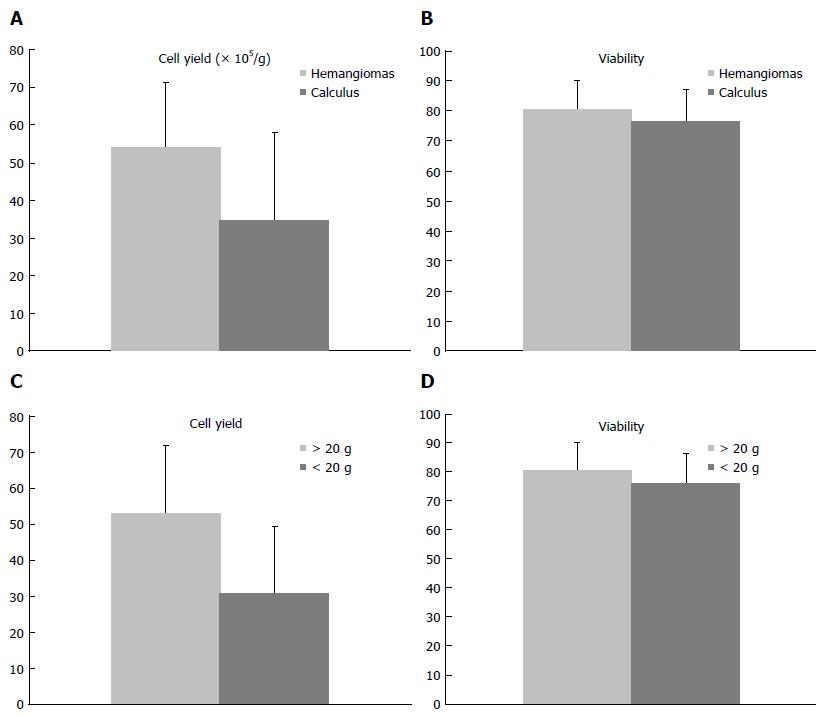
Figure 4 Samples of liver hemangioma can provide better results, in terms of cell yield, than intrahepatic duct calculi.
In addition, the size of the tissues can affect the outcome of hepatocyte isolation.
- Citation: Meng FY, Liu L, Liu J, Li CY, Wang JP, Yang FH, Chen ZS, Zhou P. Hepatocyte isolation from resected benign tissues: Results of a 5-year experience. World J Gastroenterol 2016; 22(36): 8178-8186
- URL: https://www.wjgnet.com/1007-9327/full/v22/i36/8178.htm
- DOI: https://dx.doi.org/10.3748/wjg.v22.i36.8178


