Copyright
©The Author(s) 2016.
World J Gastroenterol. Jul 21, 2016; 22(27): 6235-6245
Published online Jul 21, 2016. doi: 10.3748/wjg.v22.i27.6235
Published online Jul 21, 2016. doi: 10.3748/wjg.v22.i27.6235
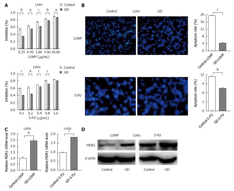
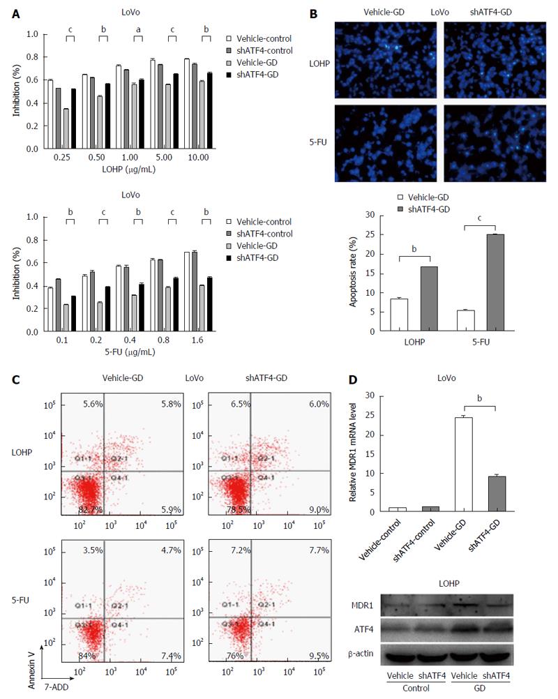
Figure 1 Glucose deprivation promotes drug resistance of colorectal cancer cells.
A: GD decreased drug susceptibility to CRC cells. LoVo cells were treated with the indicated doses of the different drugs for 48 h under GD or normal culture condition. The in vitro drug sensitivity was tested by the CCK-8 assay; B: GD inhibited LOHP- and 5-FU-induced apoptosis. LoVo cells were treated with 0.1 μg/mL LOHP or 0.05 μg/mL 5-FU for 48 h. Hoechst 33258 nuclear staining and Annexin V/7-AAD staining assays were performed to detect apoptosis. C and D: GD promoted the expression of resistance gene MDR1. The mRNA and protein levels of MDR1 were examined by qRT-PCR and Western blot, respectively. aP < 0.05, bP < 0.01,cP < 0.001, control vs GD. GD: Glucose deprivation; CRC: Colorectal cancer.
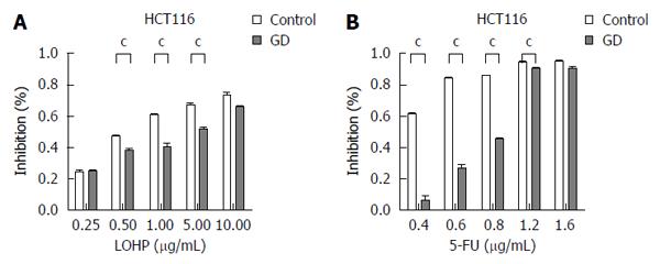
Figure 2 Glucose deprivation promotes drug resistance of HCT116 cells to LOHP and 5-FU.
HCT116 cells were treated with the indicated doses of the different drugs for 48 h under GD or the normal condition. The in vitro drug sensitivity was tested by the CCK-8 assay. cP < 0.001, control vs GD. GD: Glucose deprivation.
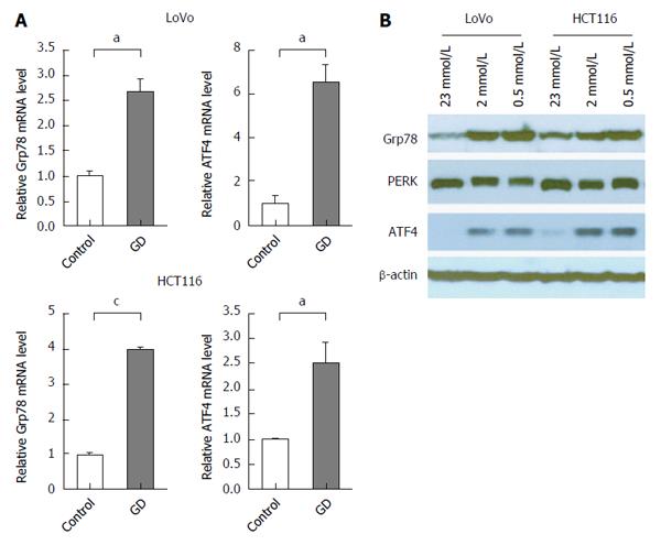
Figure 3 Grp78/PERK/ATF4 pathway is activated in glucose deprivation.
A and B: GD promoted the expression of genes involved in UPR. The mRNA and protein expression were examined by qRT-PCR and Western blot, respectively, and β-actin was used as an internal control. The phosphorylation of PERK (upward shift in the bands) indicated its activation in GD-treated CRC cells. aP < 0.05, cP < 0.001, control vs GD. GD: Glucose deprivation; CRC: Colorectal cancer.
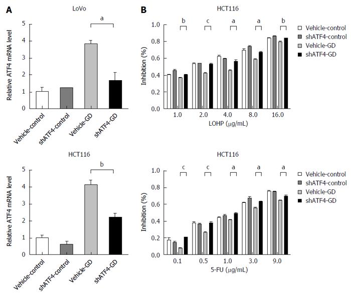
Figure 4 Down-regulation of activating transcription factor 4 significantly reverses the glucose deprivation-induced resistance of HCT116 cells to chemotherapy.
A: Silencing ATF4 expression using shATF4 in GD-treated LoVo and HCT116 cells; B: Depletion of ATF4 enhanced the sensitivity of HCT116 cells to LOHP and 5-FU. The in vitro drug sensitivity was tested using the CCK-8 assay. aP < 0.05, bP < 0.01, cP < 0.001, control vs GD. GD: Glucose deprivation.
Figure 5 Down-regulation of activating transcription factor 4 significantly reverses the glucose deprivation-induced resistance of colorectal cancer cells to chemotherapy.
A: Depletion of ATF4 enhanced the sensitivity of CRC cells to chemical drugs. Vector and shATF4 stably transfected LoVo cells were treated with the indicated doses of the different drugs for 48 h. In vitro drug sensitivity was tested using the CCK-8 assay; B and C: The apoptotic rates were much higher in the ATF4-depleted cells than in the control cells. Hoechst 33258 nuclear staining and Annexin V/7-AAD staining assays were performed to detect apoptosis; D: Depletion of ATF4 by shRNA in CRC cells led to significantly reduced expression of MDR1. The mRNA and protein levels of MDR1 were detected by qRT-PCR and Western blot, respectively, and β-actin was used as an internal controls. aP < 0.05, bP < 0.01, cP < 0.001, vehicle GD vs shATF4-GD. GD: Glucose deprivation; CRC: Colorectal cancer.
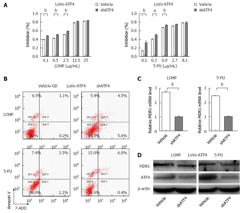
Figure 6 Inhibition of activating transcription factor 4 activity reintroduces drug sensitivity in activating transcription factor 4-overexpressing colorectal cancer cells.
A: After transfection with shATF4 or vector, ATF4-overexpressing cells were exposed to the indicated doses of LOHP or 5-FU for 48 h. Cell viabilities were determined by the CCK-8 assay; B: The apoptotic rates were much higher in the shATF4-transfected cells than in the control cells. Annexin V/7-AAD staining assay was performed to detect apoptosis; C and D: Inhibition of ATF4 decreased the MDR1 expression. The mRNA and protein levels of MDR1 in the ATF4-depleted cells were examined by qRT-PCR and Western blot, respectively, and β-actin was used as an internal controls. aP < 0.05, bP < 0.01, vehicle vs shATF4. GD: Glucose deprivation; CRC: Colorectal cancer.
Figure 7 Inhibition of activating transcription factor 4 activity reintroduces drug sensitivity in activating transcription factor 4-overexpressing HCT116 cells.
A: ATF4 knockdown increased sensitivity of HCT116-ATF4 cell to chemotherapy. The in vitro drug sensitivity was tested using the CCK-8 assay; B: ATF4 knockdown counteracted drug-induced apoptosis. The apoptotic rates were much higher in the shATF4-treated HCT116-ATF4 cells than in the control cells. aP < 0.05, bP < 0.01, cP < 0.001, vehicle vs shATF4.
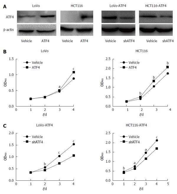
Figure 8 Activating transcription factor 4 promotes the proliferation of colorectal cancer cells.
A: We overexpressed ATF4 in LoVo and HCT116 cells and then inhibited ATF4 in these cells or their control cells, respectively; B: ATF4 overexpression enhanced the cell growth of LoVo and HCT116 cells; C: Silencing ATF4 expression inhibited the proliferation of ATF4-overexpressing cells in a dose- and time-dependent manner in LoVo and HCT116 cells. The CCK-8 assay was used to determine the cell growth rate. aP < 0.05, bP < 0.01, cP < 0.001, control vs ATF4. GD: Glucose deprivation; CRC: Colorectal cancer.
- Citation: Hu YL, Yin Y, Liu HY, Feng YY, Bian ZH, Zhou LY, Zhang JW, Fei BJ, Wang YG, Huang ZH. Glucose deprivation induces chemoresistance in colorectal cancer cells by increasing ATF4 expression. World J Gastroenterol 2016; 22(27): 6235-6245
- URL: https://www.wjgnet.com/1007-9327/full/v22/i27/6235.htm
- DOI: https://dx.doi.org/10.3748/wjg.v22.i27.6235


